Iron in PDB 5ex9: Structure of P450 Staf From Glycopeptide Antibiotic A47934 Biosynthesis; Glycerol Cryo
Protein crystallography data
The structure of Structure of P450 Staf From Glycopeptide Antibiotic A47934 Biosynthesis; Glycerol Cryo, PDB code: 5ex9
was solved by
M.J.Cryle,
V.Ulrich,
with X-Ray Crystallography technique. A brief refinement statistics is given in the table below:
| Resolution Low / High (Å) | 47.51 / 2.20 |
| Space group | P 31 2 1 |
| Cell size a, b, c (Å), α, β, γ (°) | 109.700, 109.700, 93.900, 90.00, 90.00, 120.00 |
| R / Rfree (%) | 19.1 / 22 |
Iron Binding Sites:
The binding sites of Iron atom in the Structure of P450 Staf From Glycopeptide Antibiotic A47934 Biosynthesis; Glycerol Cryo
(pdb code 5ex9). This binding sites where shown within
5.0 Angstroms radius around Iron atom.
In total only one binding site of Iron was determined in the Structure of P450 Staf From Glycopeptide Antibiotic A47934 Biosynthesis; Glycerol Cryo, PDB code: 5ex9:
In total only one binding site of Iron was determined in the Structure of P450 Staf From Glycopeptide Antibiotic A47934 Biosynthesis; Glycerol Cryo, PDB code: 5ex9:
Iron binding site 1 out of 1 in 5ex9
Go back to
Iron binding site 1 out
of 1 in the Structure of P450 Staf From Glycopeptide Antibiotic A47934 Biosynthesis; Glycerol Cryo
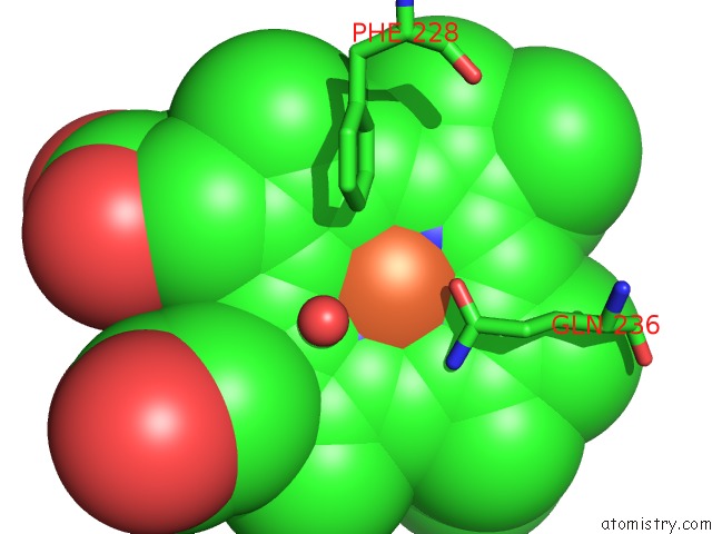
Mono view
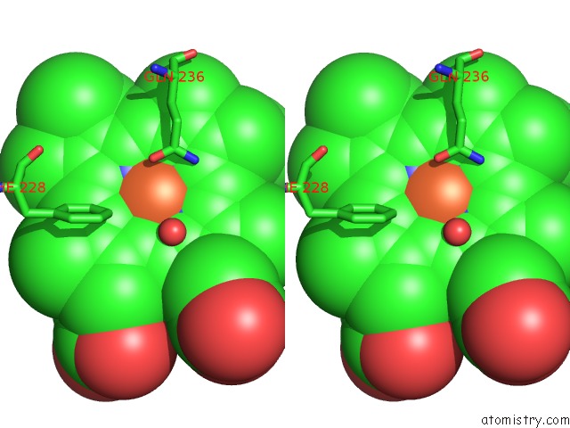
Stereo pair view
Mono view
Stereo pair view
A full contact list of Iron with other atoms in the Fe binding
site number 1 of Structure of P450 Staf From Glycopeptide Antibiotic A47934 Biosynthesis; Glycerol Cryo within 5.0Å range:
|
Reference:
V.Ulrich,
C.Brieke,
M.J.Cryle.
Biochemical and Structural Characterisation of the Second Oxidative Crosslinking Step During the Biosynthesis of the Glycopeptide Antibiotic A47934. Beilstein J Org Chem V. 12 2849 2016.
ISSN: ISSN 1860-5397
PubMed: 28144358
DOI: 10.3762/BJOC.12.284
Page generated: Tue Aug 6 00:37:01 2024
ISSN: ISSN 1860-5397
PubMed: 28144358
DOI: 10.3762/BJOC.12.284
Last articles
Zn in 9JYWZn in 9IR4
Zn in 9IR3
Zn in 9GMX
Zn in 9GMW
Zn in 9JEJ
Zn in 9ERF
Zn in 9ERE
Zn in 9EGV
Zn in 9EGW