Iron in PDB 5sx2: Crystal Structure of the D141E Mutant of B. Pseudomallei Katg at pH 8.0.
Enzymatic activity of Crystal Structure of the D141E Mutant of B. Pseudomallei Katg at pH 8.0.
All present enzymatic activity of Crystal Structure of the D141E Mutant of B. Pseudomallei Katg at pH 8.0.:
1.11.1.21;
1.11.1.21;
Protein crystallography data
The structure of Crystal Structure of the D141E Mutant of B. Pseudomallei Katg at pH 8.0., PDB code: 5sx2
was solved by
P.C.Loewen,
with X-Ray Crystallography technique. A brief refinement statistics is given in the table below:
| Resolution Low / High (Å) | 20.00 / 2.15 |
| Space group | P 21 21 21 |
| Cell size a, b, c (Å), α, β, γ (°) | 100.327, 114.824, 174.310, 90.00, 90.00, 90.00 |
| R / Rfree (%) | 14.2 / 18 |
Other elements in 5sx2:
The structure of Crystal Structure of the D141E Mutant of B. Pseudomallei Katg at pH 8.0. also contains other interesting chemical elements:
| Sodium | (Na) | 2 atoms |
Iron Binding Sites:
The binding sites of Iron atom in the Crystal Structure of the D141E Mutant of B. Pseudomallei Katg at pH 8.0.
(pdb code 5sx2). This binding sites where shown within
5.0 Angstroms radius around Iron atom.
In total 2 binding sites of Iron where determined in the Crystal Structure of the D141E Mutant of B. Pseudomallei Katg at pH 8.0., PDB code: 5sx2:
Jump to Iron binding site number: 1; 2;
In total 2 binding sites of Iron where determined in the Crystal Structure of the D141E Mutant of B. Pseudomallei Katg at pH 8.0., PDB code: 5sx2:
Jump to Iron binding site number: 1; 2;
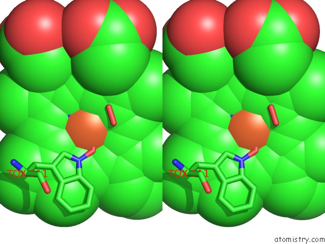
Iron binding site 1 out of 2 in 5sx2
Go back to
Iron binding site 1 out
of 2 in the Crystal Structure of the D141E Mutant of B. Pseudomallei Katg at pH 8.0.

Mono view
Stereo pair view
Mono view
Stereo pair view
A full contact list of Iron with other atoms in the Fe binding
site number 1 of Crystal Structure of the D141E Mutant of B. Pseudomallei Katg at pH 8.0. within 5.0Å range:
|
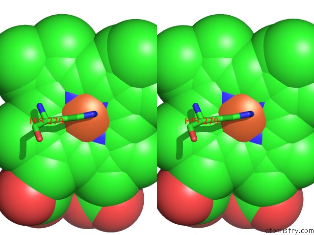
Iron binding site 2 out of 2 in 5sx2
Go back to
Iron binding site 2 out
of 2 in the Crystal Structure of the D141E Mutant of B. Pseudomallei Katg at pH 8.0.

Mono view
Stereo pair view
Mono view
Stereo pair view
A full contact list of Iron with other atoms in the Fe binding
site number 2 of Crystal Structure of the D141E Mutant of B. Pseudomallei Katg at pH 8.0. within 5.0Å range:
|
Reference:
T.Deemagarn,
B.Wiseman,
X.Carpena,
A.Ivancich,
I.Fita,
P.C.Loewen.
Two Alternative Substrate Paths For Compound I Formation and Reduction in Catalase-Peroxidase Katg From Burkholderia Pseudomallei. Proteins V. 66 219 2007.
ISSN: ESSN 1097-0134
PubMed: 17063492
Page generated: Tue Aug 6 08:48:36 2024
ISSN: ESSN 1097-0134
PubMed: 17063492
Last articles
Zn in 9J0NZn in 9J0O
Zn in 9J0P
Zn in 9FJX
Zn in 9EKB
Zn in 9C0F
Zn in 9CAH
Zn in 9CH0
Zn in 9CH3
Zn in 9CH1