Iron in PDB 5ufj: Crystal Structure of Carbonmonoxy Hemoglobin S (Liganded Sickle Cell Hemoglobin) Complexed with Gbt Compound 6
Protein crystallography data
The structure of Crystal Structure of Carbonmonoxy Hemoglobin S (Liganded Sickle Cell Hemoglobin) Complexed with Gbt Compound 6, PDB code: 5ufj
was solved by
J.R.Partridge,
R.M.Choy,
Z.Li,
B.Metcalf,
with X-Ray Crystallography technique. A brief refinement statistics is given in the table below:
Resolution Low / High (Å)
|
29.76 /
2.05
|
Space group
|
P 21 21 21
|
Cell size a, b, c (Å), α, β, γ (°)
|
56.411,
58.881,
174.386,
90.00,
90.00,
90.00
|
R / Rfree (%)
|
21.1 /
25.4
|
Iron Binding Sites:
The binding sites of Iron atom in the Crystal Structure of Carbonmonoxy Hemoglobin S (Liganded Sickle Cell Hemoglobin) Complexed with Gbt Compound 6
(pdb code 5ufj). This binding sites where shown within
5.0 Angstroms radius around Iron atom.
In total 4 binding sites of Iron where determined in the
Crystal Structure of Carbonmonoxy Hemoglobin S (Liganded Sickle Cell Hemoglobin) Complexed with Gbt Compound 6, PDB code: 5ufj:
Jump to Iron binding site number:
1;
2;
3;
4;
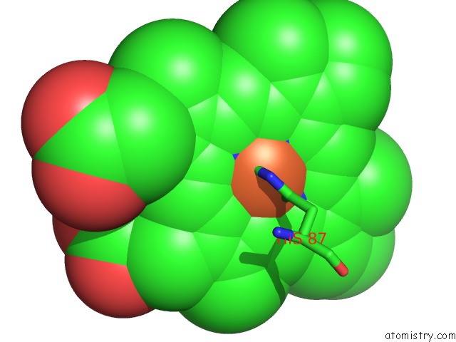
Iron binding site 1 out
of 4 in 5ufj
Go back to
Iron Binding Sites List in 5ufj
Iron binding site 1 out
of 4 in the Crystal Structure of Carbonmonoxy Hemoglobin S (Liganded Sickle Cell Hemoglobin) Complexed with Gbt Compound 6
Mono view
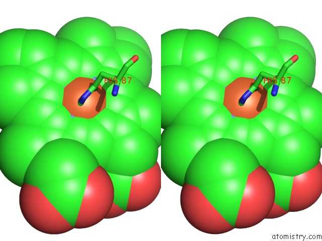
Stereo pair view
|
A full contact list of Iron with other atoms in the Fe binding
site number 1 of Crystal Structure of Carbonmonoxy Hemoglobin S (Liganded Sickle Cell Hemoglobin) Complexed with Gbt Compound 6 within 5.0Å range:
probe
|
atom
|
residue
|
distance (Å)
|
B
|
Occ
|
A:Fe201
b:68.5
occ:1.00
|
FE
|
A:HEM201
|
0.0
|
68.5
|
1.0
|
ND
|
A:HEM201
|
1.9
|
75.1
|
1.0
|
C
|
A:CMO202
|
2.0
|
0.5
|
1.0
|
NA
|
A:HEM201
|
2.0
|
71.7
|
1.0
|
NB
|
A:HEM201
|
2.0
|
58.2
|
1.0
|
NC
|
A:HEM201
|
2.1
|
58.5
|
1.0
|
NE2
|
A:HIS87
|
2.3
|
83.8
|
1.0
|
C4D
|
A:HEM201
|
2.9
|
81.0
|
1.0
|
C1D
|
A:HEM201
|
2.9
|
80.6
|
1.0
|
C1A
|
A:HEM201
|
3.0
|
78.4
|
1.0
|
C4C
|
A:HEM201
|
3.1
|
60.0
|
1.0
|
C1B
|
A:HEM201
|
3.1
|
67.1
|
1.0
|
C4B
|
A:HEM201
|
3.1
|
53.5
|
1.0
|
O
|
A:CMO202
|
3.1
|
0.4
|
1.0
|
C4A
|
A:HEM201
|
3.1
|
72.9
|
1.0
|
C1C
|
A:HEM201
|
3.1
|
53.7
|
1.0
|
CE1
|
A:HIS87
|
3.2
|
82.9
|
1.0
|
CD2
|
A:HIS87
|
3.3
|
68.7
|
1.0
|
CHA
|
A:HEM201
|
3.3
|
80.3
|
1.0
|
CHD
|
A:HEM201
|
3.4
|
64.8
|
1.0
|
CHB
|
A:HEM201
|
3.5
|
81.0
|
1.0
|
CHC
|
A:HEM201
|
3.5
|
52.2
|
1.0
|
C3D
|
A:HEM201
|
4.1
|
78.3
|
1.0
|
C2D
|
A:HEM201
|
4.1
|
74.8
|
1.0
|
C2A
|
A:HEM201
|
4.2
|
78.9
|
1.0
|
C3B
|
A:HEM201
|
4.3
|
55.6
|
1.0
|
C2B
|
A:HEM201
|
4.3
|
66.4
|
1.0
|
C3A
|
A:HEM201
|
4.3
|
81.9
|
1.0
|
C3C
|
A:HEM201
|
4.3
|
56.1
|
1.0
|
ND1
|
A:HIS87
|
4.3
|
80.2
|
1.0
|
C2C
|
A:HEM201
|
4.4
|
52.2
|
1.0
|
CG
|
A:HIS87
|
4.4
|
72.5
|
1.0
|
NE2
|
A:HIS58
|
4.4
|
0.2
|
1.0
|
CE1
|
A:HIS58
|
4.6
|
0.1
|
1.0
|
|
Iron binding site 2 out
of 4 in 5ufj
Go back to
Iron Binding Sites List in 5ufj
Iron binding site 2 out
of 4 in the Crystal Structure of Carbonmonoxy Hemoglobin S (Liganded Sickle Cell Hemoglobin) Complexed with Gbt Compound 6
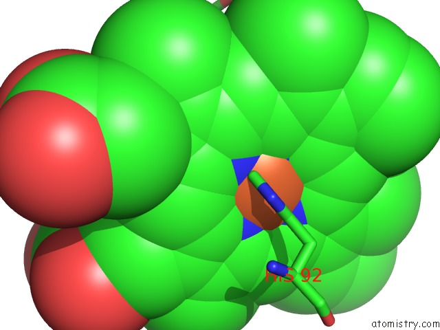
Mono view
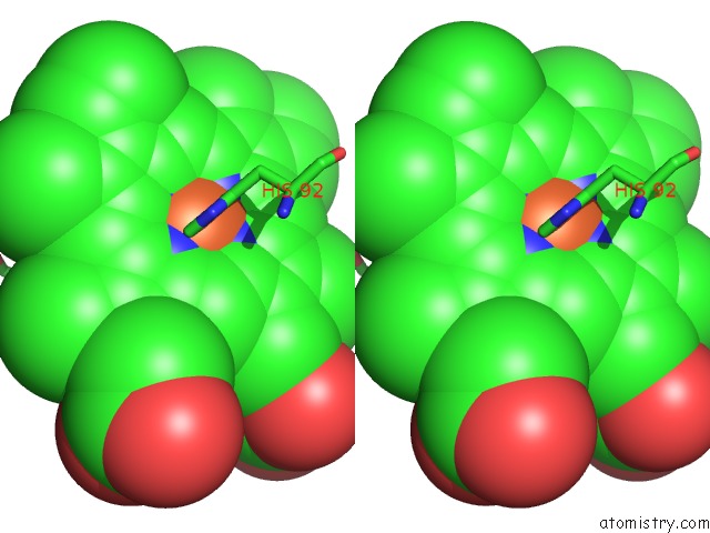
Stereo pair view
|
A full contact list of Iron with other atoms in the Fe binding
site number 2 of Crystal Structure of Carbonmonoxy Hemoglobin S (Liganded Sickle Cell Hemoglobin) Complexed with Gbt Compound 6 within 5.0Å range:
probe
|
atom
|
residue
|
distance (Å)
|
B
|
Occ
|
B:Fe201
b:64.0
occ:1.00
|
FE
|
B:HEM201
|
0.0
|
64.0
|
1.0
|
C
|
B:CMO202
|
1.6
|
0.8
|
1.0
|
NC
|
B:HEM201
|
2.0
|
70.7
|
1.0
|
ND
|
B:HEM201
|
2.0
|
74.0
|
1.0
|
NA
|
B:HEM201
|
2.0
|
79.4
|
1.0
|
NB
|
B:HEM201
|
2.2
|
78.1
|
1.0
|
NE2
|
B:HIS92
|
2.5
|
63.3
|
1.0
|
O
|
B:CMO202
|
2.6
|
0.2
|
1.0
|
C4C
|
B:HEM201
|
3.0
|
66.5
|
1.0
|
C4D
|
B:HEM201
|
3.0
|
82.5
|
1.0
|
C1D
|
B:HEM201
|
3.0
|
72.9
|
1.0
|
C1A
|
B:HEM201
|
3.0
|
81.3
|
1.0
|
C1C
|
B:HEM201
|
3.1
|
67.1
|
1.0
|
C4A
|
B:HEM201
|
3.1
|
86.9
|
1.0
|
C1B
|
B:HEM201
|
3.2
|
79.6
|
1.0
|
CD2
|
B:HIS92
|
3.2
|
68.2
|
1.0
|
C4B
|
B:HEM201
|
3.2
|
74.2
|
1.0
|
CHD
|
B:HEM201
|
3.4
|
74.1
|
1.0
|
CHA
|
B:HEM201
|
3.4
|
82.5
|
1.0
|
CHB
|
B:HEM201
|
3.5
|
86.2
|
1.0
|
CHC
|
B:HEM201
|
3.5
|
66.8
|
1.0
|
CE1
|
B:HIS92
|
3.6
|
77.5
|
1.0
|
C3C
|
B:HEM201
|
4.2
|
65.4
|
1.0
|
C3D
|
B:HEM201
|
4.2
|
88.3
|
1.0
|
C2C
|
B:HEM201
|
4.2
|
68.1
|
1.0
|
C2D
|
B:HEM201
|
4.2
|
78.7
|
1.0
|
C2A
|
B:HEM201
|
4.3
|
85.7
|
1.0
|
NE2
|
B:HIS63
|
4.3
|
88.4
|
1.0
|
C3A
|
B:HEM201
|
4.3
|
83.4
|
1.0
|
C2B
|
B:HEM201
|
4.4
|
82.0
|
1.0
|
C3B
|
B:HEM201
|
4.4
|
75.9
|
1.0
|
CG
|
B:HIS92
|
4.4
|
78.0
|
1.0
|
ND1
|
B:HIS92
|
4.6
|
82.6
|
1.0
|
CE1
|
B:HIS63
|
4.7
|
94.7
|
1.0
|
CG2
|
B:VAL67
|
4.9
|
72.3
|
1.0
|
|
Iron binding site 3 out
of 4 in 5ufj
Go back to
Iron Binding Sites List in 5ufj
Iron binding site 3 out
of 4 in the Crystal Structure of Carbonmonoxy Hemoglobin S (Liganded Sickle Cell Hemoglobin) Complexed with Gbt Compound 6
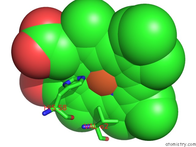
Mono view
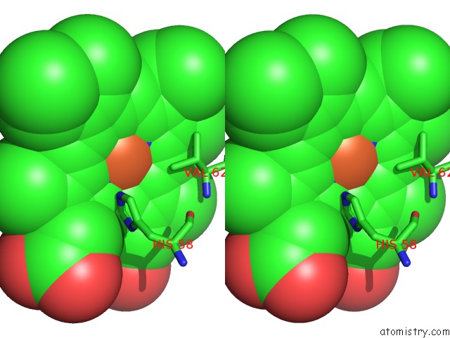
Stereo pair view
|
A full contact list of Iron with other atoms in the Fe binding
site number 3 of Crystal Structure of Carbonmonoxy Hemoglobin S (Liganded Sickle Cell Hemoglobin) Complexed with Gbt Compound 6 within 5.0Å range:
probe
|
atom
|
residue
|
distance (Å)
|
B
|
Occ
|
C:Fe201
b:35.4
occ:1.00
|
FE
|
C:HEM201
|
0.0
|
35.4
|
1.0
|
C
|
C:CMO202
|
1.8
|
0.1
|
1.0
|
ND
|
C:HEM201
|
2.0
|
45.9
|
1.0
|
NB
|
C:HEM201
|
2.0
|
34.0
|
1.0
|
NC
|
C:HEM201
|
2.0
|
33.2
|
1.0
|
NA
|
C:HEM201
|
2.1
|
38.1
|
1.0
|
NE2
|
C:HIS87
|
2.2
|
33.6
|
1.0
|
C1D
|
C:HEM201
|
2.9
|
39.6
|
1.0
|
C4C
|
C:HEM201
|
3.0
|
35.0
|
1.0
|
C4A
|
C:HEM201
|
3.0
|
41.0
|
1.0
|
C1B
|
C:HEM201
|
3.0
|
34.7
|
1.0
|
O
|
C:CMO202
|
3.0
|
0.7
|
1.0
|
C4D
|
C:HEM201
|
3.1
|
50.7
|
1.0
|
CE1
|
C:HIS87
|
3.1
|
42.8
|
1.0
|
C4B
|
C:HEM201
|
3.1
|
40.2
|
1.0
|
C1C
|
C:HEM201
|
3.1
|
35.3
|
1.0
|
C1A
|
C:HEM201
|
3.1
|
53.2
|
1.0
|
CD2
|
C:HIS87
|
3.2
|
42.5
|
1.0
|
CHD
|
C:HEM201
|
3.3
|
37.1
|
1.0
|
CHB
|
C:HEM201
|
3.3
|
35.4
|
1.0
|
CHC
|
C:HEM201
|
3.5
|
41.4
|
1.0
|
CHA
|
C:HEM201
|
3.5
|
46.5
|
1.0
|
C2D
|
C:HEM201
|
4.2
|
44.3
|
1.0
|
C3C
|
C:HEM201
|
4.2
|
41.2
|
1.0
|
ND1
|
C:HIS87
|
4.2
|
35.1
|
1.0
|
C3D
|
C:HEM201
|
4.2
|
55.8
|
1.0
|
NE2
|
C:HIS58
|
4.2
|
60.1
|
1.0
|
C3A
|
C:HEM201
|
4.2
|
41.5
|
1.0
|
C2B
|
C:HEM201
|
4.2
|
35.8
|
1.0
|
C3B
|
C:HEM201
|
4.3
|
31.3
|
1.0
|
C2C
|
C:HEM201
|
4.3
|
36.0
|
1.0
|
CG
|
C:HIS87
|
4.3
|
40.5
|
1.0
|
C2A
|
C:HEM201
|
4.3
|
42.3
|
1.0
|
CE1
|
C:HIS58
|
4.6
|
47.3
|
1.0
|
CG2
|
C:VAL62
|
4.8
|
39.6
|
1.0
|
|
Iron binding site 4 out
of 4 in 5ufj
Go back to
Iron Binding Sites List in 5ufj
Iron binding site 4 out
of 4 in the Crystal Structure of Carbonmonoxy Hemoglobin S (Liganded Sickle Cell Hemoglobin) Complexed with Gbt Compound 6
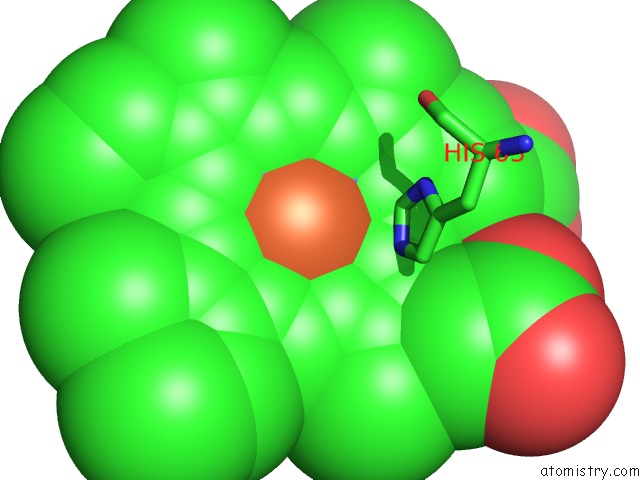
Mono view
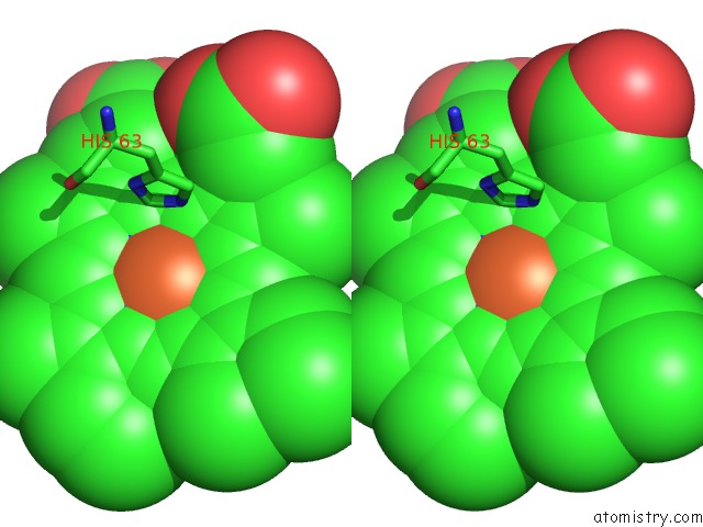
Stereo pair view
|
A full contact list of Iron with other atoms in the Fe binding
site number 4 of Crystal Structure of Carbonmonoxy Hemoglobin S (Liganded Sickle Cell Hemoglobin) Complexed with Gbt Compound 6 within 5.0Å range:
probe
|
atom
|
residue
|
distance (Å)
|
B
|
Occ
|
D:Fe201
b:90.3
occ:1.00
|
FE
|
D:HEM201
|
0.0
|
90.3
|
1.0
|
C
|
D:CMO202
|
1.9
|
0.3
|
1.0
|
NB
|
D:HEM201
|
2.0
|
87.2
|
1.0
|
ND
|
D:HEM201
|
2.0
|
94.4
|
1.0
|
NC
|
D:HEM201
|
2.0
|
80.9
|
1.0
|
NA
|
D:HEM201
|
2.1
|
0.8
|
1.0
|
NE2
|
D:HIS92
|
2.4
|
93.8
|
1.0
|
C4B
|
D:HEM201
|
3.0
|
80.5
|
1.0
|
O
|
D:CMO202
|
3.0
|
0.4
|
1.0
|
C1C
|
D:HEM201
|
3.0
|
75.4
|
1.0
|
C4D
|
D:HEM201
|
3.0
|
0.0
|
1.0
|
C1A
|
D:HEM201
|
3.1
|
0.4
|
1.0
|
C4C
|
D:HEM201
|
3.1
|
79.5
|
1.0
|
CE1
|
D:HIS92
|
3.1
|
93.2
|
1.0
|
C1D
|
D:HEM201
|
3.1
|
91.2
|
1.0
|
C1B
|
D:HEM201
|
3.1
|
91.4
|
1.0
|
C4A
|
D:HEM201
|
3.1
|
0.2
|
1.0
|
CD2
|
D:HIS92
|
3.3
|
92.8
|
1.0
|
CHC
|
D:HEM201
|
3.3
|
75.3
|
1.0
|
CHA
|
D:HEM201
|
3.4
|
0.8
|
1.0
|
CHD
|
D:HEM201
|
3.5
|
84.2
|
1.0
|
CHB
|
D:HEM201
|
3.5
|
98.9
|
1.0
|
ND1
|
D:HIS92
|
4.1
|
96.7
|
1.0
|
C3B
|
D:HEM201
|
4.2
|
80.7
|
1.0
|
C2C
|
D:HEM201
|
4.2
|
72.1
|
1.0
|
CG
|
D:HIS92
|
4.2
|
92.8
|
1.0
|
C3D
|
D:HEM201
|
4.2
|
0.5
|
1.0
|
C3C
|
D:HEM201
|
4.3
|
73.0
|
1.0
|
C2B
|
D:HEM201
|
4.3
|
87.1
|
1.0
|
C2D
|
D:HEM201
|
4.3
|
97.0
|
1.0
|
C2A
|
D:HEM201
|
4.3
|
0.4
|
1.0
|
C3A
|
D:HEM201
|
4.3
|
0.8
|
1.0
|
CE1
|
D:HIS63
|
4.4
|
0.9
|
1.0
|
NE2
|
D:HIS63
|
4.4
|
0.4
|
1.0
|
|
Reference:
B.Metcalf,
C.Chuang,
K.Dufu,
M.P.Patel,
A.Silva-Garcia,
C.Johnson,
Q.Lu,
J.R.Partridge,
L.Patskovska,
Y.Patskovsky,
S.C.Almo,
M.P.Jacobson,
L.Hua,
Q.Xu,
S.L.Gwaltney,
C.Yee,
J.Harris,
B.P.Morgan,
J.James,
D.Xu,
A.Hutchaleelaha,
K.Paulvannan,
D.Oksenberg,
Z.Li.
Discovery of GBT440, An Orally Bioavailable R-State Stabilizer of Sickle Cell Hemoglobin. Acs Med Chem Lett V. 8 321 2017.
ISSN: ISSN 1948-5875
PubMed: 28337324
DOI: 10.1021/ACSMEDCHEMLETT.6B00491
Page generated: Tue Aug 6 09:52:40 2024
|