Iron in PDB 5ve3: Crystal Structure of Wild-Type Persulfide Dioxygenase-Rhodanese Fusion Protein From Burkholderia Phytofirmans
Enzymatic activity of Crystal Structure of Wild-Type Persulfide Dioxygenase-Rhodanese Fusion Protein From Burkholderia Phytofirmans
All present enzymatic activity of Crystal Structure of Wild-Type Persulfide Dioxygenase-Rhodanese Fusion Protein From Burkholderia Phytofirmans:
1.13.11.18; 2.8.1.1;
1.13.11.18; 2.8.1.1;
Protein crystallography data
The structure of Crystal Structure of Wild-Type Persulfide Dioxygenase-Rhodanese Fusion Protein From Burkholderia Phytofirmans, PDB code: 5ve3
was solved by
N.Motl,
M.A.Skiba,
J.L.Smith,
R.Banerjee,
with X-Ray Crystallography technique. A brief refinement statistics is given in the table below:
| Resolution Low / High (Å) | 43.60 / 1.79 |
| Space group | P 21 21 21 |
| Cell size a, b, c (Å), α, β, γ (°) | 63.689, 108.333, 119.616, 90.00, 90.00, 90.00 |
| R / Rfree (%) | 16.1 / 19.6 |
Iron Binding Sites:
The binding sites of Iron atom in the Crystal Structure of Wild-Type Persulfide Dioxygenase-Rhodanese Fusion Protein From Burkholderia Phytofirmans
(pdb code 5ve3). This binding sites where shown within
5.0 Angstroms radius around Iron atom.
In total 2 binding sites of Iron where determined in the Crystal Structure of Wild-Type Persulfide Dioxygenase-Rhodanese Fusion Protein From Burkholderia Phytofirmans, PDB code: 5ve3:
Jump to Iron binding site number: 1; 2;
In total 2 binding sites of Iron where determined in the Crystal Structure of Wild-Type Persulfide Dioxygenase-Rhodanese Fusion Protein From Burkholderia Phytofirmans, PDB code: 5ve3:
Jump to Iron binding site number: 1; 2;
Iron binding site 1 out of 2 in 5ve3
Go back to
Iron binding site 1 out
of 2 in the Crystal Structure of Wild-Type Persulfide Dioxygenase-Rhodanese Fusion Protein From Burkholderia Phytofirmans
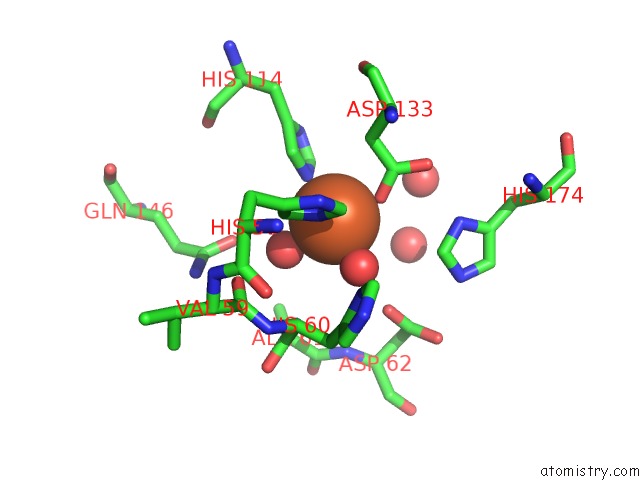
Mono view
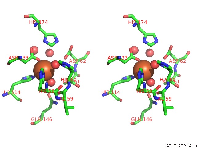
Stereo pair view
Mono view
Stereo pair view
A full contact list of Iron with other atoms in the Fe binding
site number 1 of Crystal Structure of Wild-Type Persulfide Dioxygenase-Rhodanese Fusion Protein From Burkholderia Phytofirmans within 5.0Å range:
|
Iron binding site 2 out of 2 in 5ve3
Go back to
Iron binding site 2 out
of 2 in the Crystal Structure of Wild-Type Persulfide Dioxygenase-Rhodanese Fusion Protein From Burkholderia Phytofirmans
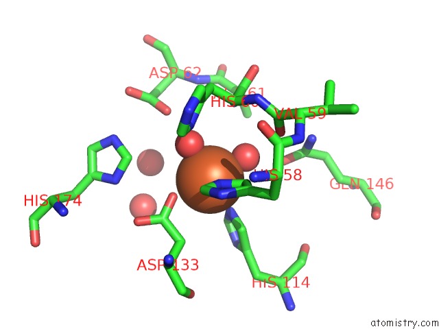
Mono view
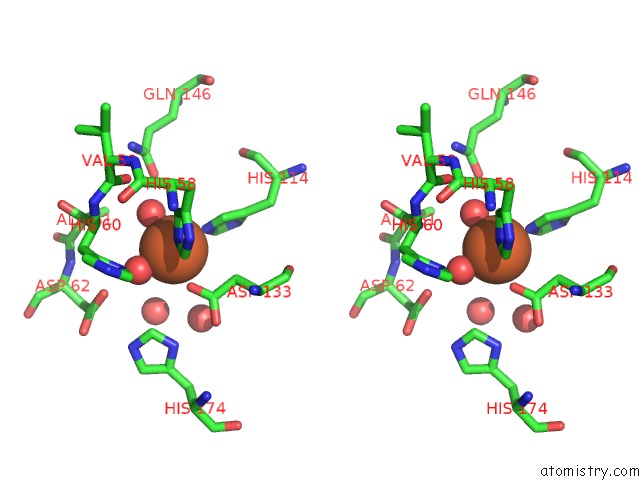
Stereo pair view
Mono view
Stereo pair view
A full contact list of Iron with other atoms in the Fe binding
site number 2 of Crystal Structure of Wild-Type Persulfide Dioxygenase-Rhodanese Fusion Protein From Burkholderia Phytofirmans within 5.0Å range:
|
Reference:
N.Motl,
M.A.Skiba,
O.Kabil,
J.L.Smith,
R.Banerjee.
Structural and Biochemical Analyses Indicate That A Bacterial Persulfide Dioxygenase-Rhodanese Fusion Protein Functions in Sulfur Assimilation. J. Biol. Chem. V. 292 14026 2017.
ISSN: ESSN 1083-351X
PubMed: 28684420
DOI: 10.1074/JBC.M117.790170
Page generated: Tue Aug 6 10:26:26 2024
ISSN: ESSN 1083-351X
PubMed: 28684420
DOI: 10.1074/JBC.M117.790170
Last articles
Zn in 9J0NZn in 9J0O
Zn in 9J0P
Zn in 9FJX
Zn in 9EKB
Zn in 9C0F
Zn in 9CAH
Zn in 9CH0
Zn in 9CH3
Zn in 9CH1