Iron in PDB 6kbh: Crystal Structure of An Intact Type IV Self-Sufficient Cytochrome P450 Monooxygenase
Protein crystallography data
The structure of Crystal Structure of An Intact Type IV Self-Sufficient Cytochrome P450 Monooxygenase, PDB code: 6kbh
was solved by
R.Gong,
L.J.Wu,
Y.Zhang,
Z.Liu,
S.Dou,
R.G.Zhang,
J.H.Xu,
C.Tang,
J.H.Zhou,
with X-Ray Crystallography technique. A brief refinement statistics is given in the table below:
Resolution Low / High (Å)
|
46.89 /
2.60
|
Space group
|
C 1 2 1
|
Cell size a, b, c (Å), α, β, γ (°)
|
131.762,
55.484,
135.960,
90.00,
98.12,
90.00
|
R / Rfree (%)
|
23.6 /
27.7
|
Iron Binding Sites:
The binding sites of Iron atom in the Crystal Structure of An Intact Type IV Self-Sufficient Cytochrome P450 Monooxygenase
(pdb code 6kbh). This binding sites where shown within
5.0 Angstroms radius around Iron atom.
In total 3 binding sites of Iron where determined in the
Crystal Structure of An Intact Type IV Self-Sufficient Cytochrome P450 Monooxygenase, PDB code: 6kbh:
Jump to Iron binding site number:
1;
2;
3;
Iron binding site 1 out
of 3 in 6kbh
Go back to
Iron Binding Sites List in 6kbh
Iron binding site 1 out
of 3 in the Crystal Structure of An Intact Type IV Self-Sufficient Cytochrome P450 Monooxygenase
Mono view
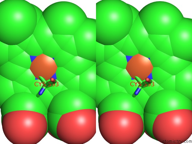
Stereo pair view
|
A full contact list of Iron with other atoms in the Fe binding
site number 1 of Crystal Structure of An Intact Type IV Self-Sufficient Cytochrome P450 Monooxygenase within 5.0Å range:
probe
|
atom
|
residue
|
distance (Å)
|
B
|
Occ
|
A:Fe802
b:75.7
occ:1.00
|
FE
|
A:HEM802
|
0.0
|
75.7
|
1.0
|
NB
|
A:HEM802
|
1.9
|
67.4
|
1.0
|
NC
|
A:HEM802
|
2.0
|
75.2
|
1.0
|
NA
|
A:HEM802
|
2.0
|
70.3
|
1.0
|
ND
|
A:HEM802
|
2.0
|
64.8
|
1.0
|
SG
|
A:CYS393
|
2.3
|
63.0
|
1.0
|
C4A
|
A:HEM802
|
2.9
|
54.1
|
1.0
|
C1C
|
A:HEM802
|
2.9
|
75.7
|
1.0
|
C1B
|
A:HEM802
|
3.0
|
62.9
|
1.0
|
C4B
|
A:HEM802
|
3.1
|
64.0
|
1.0
|
C4C
|
A:HEM802
|
3.1
|
70.3
|
1.0
|
C1A
|
A:HEM802
|
3.1
|
72.7
|
1.0
|
C1D
|
A:HEM802
|
3.2
|
63.4
|
1.0
|
O
|
A:HOH906
|
3.2
|
64.2
|
1.0
|
C4D
|
A:HEM802
|
3.2
|
67.0
|
1.0
|
CB
|
A:CYS393
|
3.3
|
61.0
|
1.0
|
CHB
|
A:HEM802
|
3.5
|
63.9
|
1.0
|
CHC
|
A:HEM802
|
3.5
|
71.4
|
1.0
|
CHD
|
A:HEM802
|
3.8
|
75.6
|
1.0
|
CHA
|
A:HEM802
|
3.8
|
72.5
|
1.0
|
O
|
A:ALA282
|
3.9
|
75.9
|
1.0
|
CA
|
A:CYS393
|
4.1
|
66.2
|
1.0
|
C2C
|
A:HEM802
|
4.2
|
69.6
|
1.0
|
C3A
|
A:HEM802
|
4.3
|
62.3
|
1.0
|
C3C
|
A:HEM802
|
4.3
|
75.0
|
1.0
|
C2B
|
A:HEM802
|
4.3
|
72.7
|
1.0
|
C3B
|
A:HEM802
|
4.3
|
59.5
|
1.0
|
C2A
|
A:HEM802
|
4.4
|
58.5
|
1.0
|
C2D
|
A:HEM802
|
4.5
|
60.7
|
1.0
|
C3D
|
A:HEM802
|
4.5
|
66.2
|
1.0
|
CB
|
A:ALA282
|
4.8
|
75.0
|
1.0
|
C
|
A:ALA282
|
4.9
|
75.3
|
1.0
|
C
|
A:CYS393
|
4.9
|
71.3
|
1.0
|
|
Iron binding site 2 out
of 3 in 6kbh
Go back to
Iron Binding Sites List in 6kbh
Iron binding site 2 out
of 3 in the Crystal Structure of An Intact Type IV Self-Sufficient Cytochrome P450 Monooxygenase
Mono view
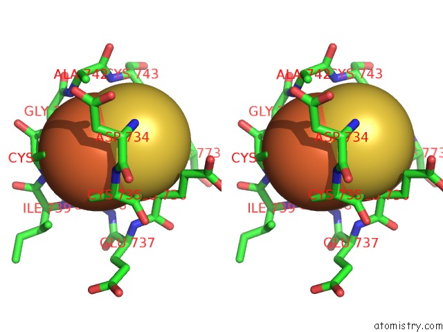
Stereo pair view
|
A full contact list of Iron with other atoms in the Fe binding
site number 2 of Crystal Structure of An Intact Type IV Self-Sufficient Cytochrome P450 Monooxygenase within 5.0Å range:
probe
|
atom
|
residue
|
distance (Å)
|
B
|
Occ
|
A:Fe803
b:84.3
occ:1.00
|
FE1
|
A:FES803
|
0.0
|
84.3
|
1.0
|
S1
|
A:FES803
|
2.1
|
82.0
|
1.0
|
S2
|
A:FES803
|
2.1
|
82.0
|
1.0
|
SG
|
A:CYS740
|
2.3
|
92.6
|
1.0
|
SG
|
A:CYS735
|
2.3
|
95.0
|
1.0
|
FE2
|
A:FES803
|
3.0
|
83.4
|
1.0
|
N
|
A:GLY741
|
3.4
|
89.2
|
1.0
|
CB
|
A:CYS740
|
3.4
|
95.8
|
1.0
|
N
|
A:CYS740
|
3.5
|
82.8
|
1.0
|
CB
|
A:CYS735
|
3.5
|
92.4
|
1.0
|
N
|
A:GLU736
|
3.6
|
99.2
|
1.0
|
CA
|
A:CYS740
|
3.8
|
86.6
|
1.0
|
N
|
A:CYS735
|
3.8
|
0.2
|
1.0
|
N
|
A:ALA742
|
3.9
|
96.8
|
1.0
|
C
|
A:CYS740
|
4.0
|
93.6
|
1.0
|
CA
|
A:CYS735
|
4.1
|
89.4
|
1.0
|
N
|
A:ILE739
|
4.3
|
78.8
|
1.0
|
C
|
A:CYS735
|
4.3
|
94.2
|
1.0
|
CA
|
A:GLY741
|
4.4
|
85.5
|
1.0
|
CA
|
A:GLU736
|
4.5
|
0.8
|
1.0
|
C
|
A:GLY741
|
4.5
|
93.6
|
1.0
|
C
|
A:ILE739
|
4.6
|
93.6
|
1.0
|
SG
|
A:CYS743
|
4.7
|
91.8
|
1.0
|
N
|
A:GLY738
|
4.7
|
96.0
|
1.0
|
CB
|
A:ALA742
|
4.7
|
89.5
|
1.0
|
N
|
A:CYS743
|
4.7
|
91.9
|
1.0
|
SG
|
A:CYS773
|
4.8
|
0.5
|
1.0
|
N
|
A:ASP734
|
4.8
|
0.0
|
1.0
|
CA
|
A:ALA742
|
4.8
|
95.0
|
1.0
|
C
|
A:GLY738
|
4.9
|
75.5
|
1.0
|
C
|
A:ASP734
|
5.0
|
0.7
|
1.0
|
CA
|
A:GLY738
|
5.0
|
77.2
|
1.0
|
N
|
A:GLU737
|
5.0
|
0.4
|
1.0
|
|
Iron binding site 3 out
of 3 in 6kbh
Go back to
Iron Binding Sites List in 6kbh
Iron binding site 3 out
of 3 in the Crystal Structure of An Intact Type IV Self-Sufficient Cytochrome P450 Monooxygenase
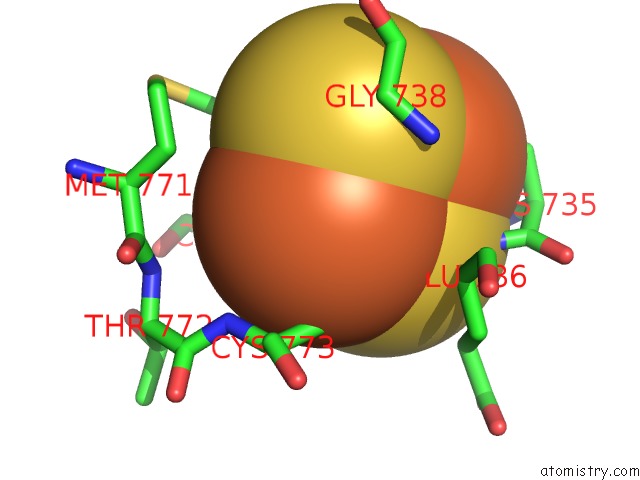
Mono view
Stereo pair view
|
A full contact list of Iron with other atoms in the Fe binding
site number 3 of Crystal Structure of An Intact Type IV Self-Sufficient Cytochrome P450 Monooxygenase within 5.0Å range:
probe
|
atom
|
residue
|
distance (Å)
|
B
|
Occ
|
A:Fe803
b:83.4
occ:1.00
|
FE2
|
A:FES803
|
0.0
|
83.4
|
1.0
|
S2
|
A:FES803
|
2.1
|
82.0
|
1.0
|
S1
|
A:FES803
|
2.1
|
82.0
|
1.0
|
SG
|
A:CYS743
|
2.3
|
91.8
|
1.0
|
SG
|
A:CYS773
|
2.3
|
0.5
|
1.0
|
FE1
|
A:FES803
|
3.0
|
84.3
|
1.0
|
CB
|
A:CYS743
|
3.3
|
84.0
|
1.0
|
CB
|
A:CYS773
|
3.3
|
97.0
|
1.0
|
N
|
A:CYS773
|
4.1
|
95.6
|
1.0
|
CA
|
A:GLU736
|
4.1
|
0.8
|
1.0
|
N
|
A:GLU736
|
4.2
|
99.2
|
1.0
|
CA
|
A:CYS773
|
4.3
|
95.2
|
1.0
|
SG
|
A:CYS735
|
4.4
|
95.0
|
1.0
|
N
|
A:CYS743
|
4.4
|
91.9
|
1.0
|
CE
|
A:MET771
|
4.4
|
84.3
|
1.0
|
CA
|
A:CYS743
|
4.4
|
83.2
|
1.0
|
N
|
A:GLY738
|
4.6
|
96.0
|
1.0
|
CA
|
A:GLY738
|
4.7
|
77.2
|
1.0
|
CB
|
A:MET771
|
4.8
|
93.8
|
1.0
|
N
|
A:THR772
|
4.9
|
99.3
|
1.0
|
C
|
A:GLU736
|
4.9
|
0.8
|
1.0
|
|
Reference:
R.Gong,
L.J.Wu,
Y.Zhang,
Z.Liu,
S.Dou,
R.G.Zhang,
J.H.Xu,
C.Tang,
J.H.Zhou.
Crystal Structure of An Intact Type IV Self-Sufficient Cytochrome P450 Monooxygenase To Be Published.
Page generated: Wed Aug 7 00:16:29 2024
|