Iron in PDB 6zzu: Partial Structure of the Substrate-Free Tyrosine Hydroxylase (Apo-Th).
Enzymatic activity of Partial Structure of the Substrate-Free Tyrosine Hydroxylase (Apo-Th).
All present enzymatic activity of Partial Structure of the Substrate-Free Tyrosine Hydroxylase (Apo-Th).:
1.14.16.2;
1.14.16.2;
Iron Binding Sites:
The binding sites of Iron atom in the Partial Structure of the Substrate-Free Tyrosine Hydroxylase (Apo-Th).
(pdb code 6zzu). This binding sites where shown within
5.0 Angstroms radius around Iron atom.
In total 4 binding sites of Iron where determined in the Partial Structure of the Substrate-Free Tyrosine Hydroxylase (Apo-Th)., PDB code: 6zzu:
Jump to Iron binding site number: 1; 2; 3; 4;
In total 4 binding sites of Iron where determined in the Partial Structure of the Substrate-Free Tyrosine Hydroxylase (Apo-Th)., PDB code: 6zzu:
Jump to Iron binding site number: 1; 2; 3; 4;
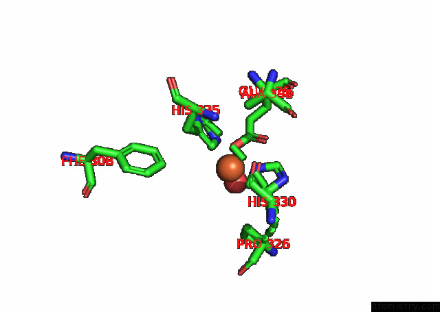
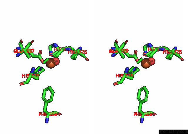
Iron binding site 1 out of 4 in 6zzu
Go back to
Iron binding site 1 out
of 4 in the Partial Structure of the Substrate-Free Tyrosine Hydroxylase (Apo-Th).
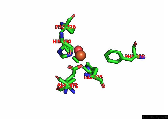
Mono view
Stereo pair view
Mono view
Stereo pair view
A full contact list of Iron with other atoms in the Fe binding
site number 1 of Partial Structure of the Substrate-Free Tyrosine Hydroxylase (Apo-Th). within 5.0Å range:
|
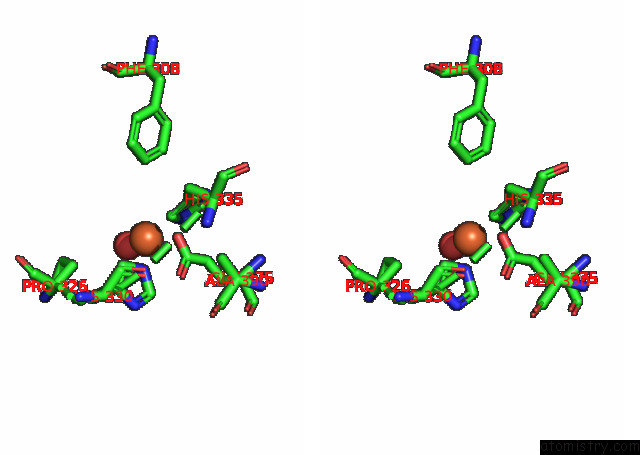
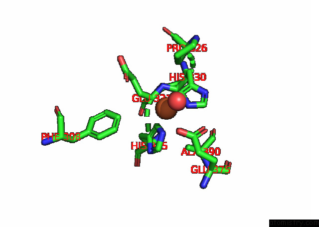
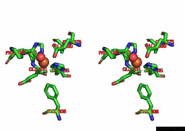
Iron binding site 2 out of 4 in 6zzu
Go back to
Iron binding site 2 out
of 4 in the Partial Structure of the Substrate-Free Tyrosine Hydroxylase (Apo-Th).
Mono view
Stereo pair view
Mono view
Stereo pair view
A full contact list of Iron with other atoms in the Fe binding
site number 2 of Partial Structure of the Substrate-Free Tyrosine Hydroxylase (Apo-Th). within 5.0Å range:
|
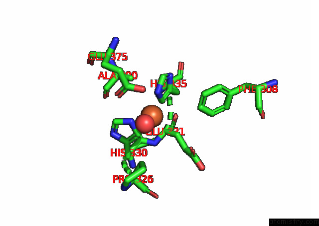
Iron binding site 3 out of 4 in 6zzu
Go back to
Iron binding site 3 out
of 4 in the Partial Structure of the Substrate-Free Tyrosine Hydroxylase (Apo-Th).
Mono view
Stereo pair view
Mono view
Stereo pair view
A full contact list of Iron with other atoms in the Fe binding
site number 3 of Partial Structure of the Substrate-Free Tyrosine Hydroxylase (Apo-Th). within 5.0Å range:
|
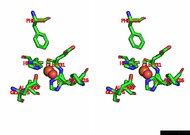
Iron binding site 4 out of 4 in 6zzu
Go back to
Iron binding site 4 out
of 4 in the Partial Structure of the Substrate-Free Tyrosine Hydroxylase (Apo-Th).
Mono view
Stereo pair view
Mono view
Stereo pair view
A full contact list of Iron with other atoms in the Fe binding
site number 4 of Partial Structure of the Substrate-Free Tyrosine Hydroxylase (Apo-Th). within 5.0Å range:
|
Reference:
M.T.Bueno-Carrasco,
J.Cuellar,
M.I.Flydal,
C.Santiago,
T.A.Krakenes,
R.Kleppe,
J.R.Lopez-Blanco,
K.Teigen,
S.Alvira,
P.Chacon,
A.Martinez,
J.M.Valpuesta.
The Structure of Human Tyrosine Hydroxylase Reveals the Mechanism For Feedback Inhibition By Dopamine To Be Published.
Page generated: Wed Aug 7 21:40:53 2024
Last articles
Zn in 9J0NZn in 9J0O
Zn in 9J0P
Zn in 9FJX
Zn in 9EKB
Zn in 9C0F
Zn in 9CAH
Zn in 9CH0
Zn in 9CH3
Zn in 9CH1