Iron in PDB 7lvz: Crystal Structure of Ado
Enzymatic activity of Crystal Structure of Ado
All present enzymatic activity of Crystal Structure of Ado:
1.13.11.19;
1.13.11.19;
Protein crystallography data
The structure of Crystal Structure of Ado, PDB code: 7lvz
was solved by
C.A.Bingman,
R.L.Fernandez,
R.W.Smith,
B.G.Fox,
T.C.Brunold,
with X-Ray Crystallography technique. A brief refinement statistics is given in the table below:
| Resolution Low / High (Å) | 39.17 / 1.89 |
| Space group | P 21 21 21 |
| Cell size a, b, c (Å), α, β, γ (°) | 54.296, 139.525, 142.007, 90, 90, 90 |
| R / Rfree (%) | 19.1 / 22.8 |
Other elements in 7lvz:
The structure of Crystal Structure of Ado also contains other interesting chemical elements:
| Chlorine | (Cl) | 4 atoms |
| Magnesium | (Mg) | 2 atoms |
Iron Binding Sites:
The binding sites of Iron atom in the Crystal Structure of Ado
(pdb code 7lvz). This binding sites where shown within
5.0 Angstroms radius around Iron atom.
In total 4 binding sites of Iron where determined in the Crystal Structure of Ado, PDB code: 7lvz:
Jump to Iron binding site number: 1; 2; 3; 4;
In total 4 binding sites of Iron where determined in the Crystal Structure of Ado, PDB code: 7lvz:
Jump to Iron binding site number: 1; 2; 3; 4;
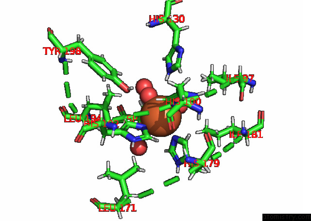
Iron binding site 1 out of 4 in 7lvz
Go back to
Iron binding site 1 out
of 4 in the Crystal Structure of Ado
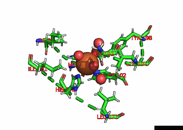
Mono view
Stereo pair view
Mono view
Stereo pair view
A full contact list of Iron with other atoms in the Fe binding
site number 1 of Crystal Structure of Ado within 5.0Å range:
|
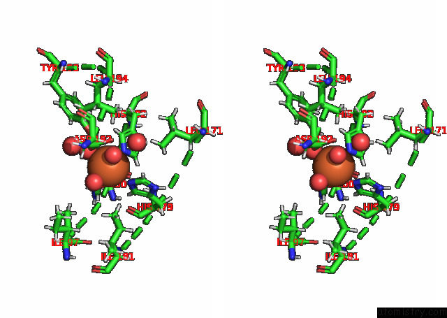
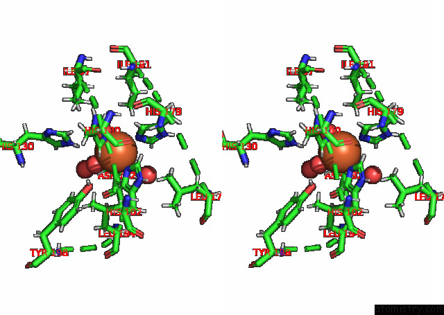
Iron binding site 2 out of 4 in 7lvz
Go back to
Iron binding site 2 out
of 4 in the Crystal Structure of Ado
Mono view
Stereo pair view
Mono view
Stereo pair view
A full contact list of Iron with other atoms in the Fe binding
site number 2 of Crystal Structure of Ado within 5.0Å range:
|
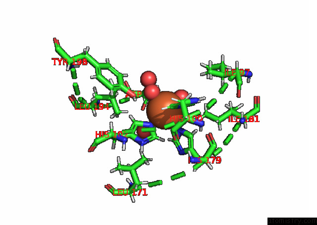
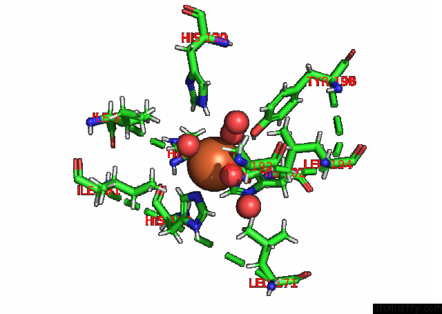
Iron binding site 3 out of 4 in 7lvz
Go back to
Iron binding site 3 out
of 4 in the Crystal Structure of Ado
Mono view
Stereo pair view
Mono view
Stereo pair view
A full contact list of Iron with other atoms in the Fe binding
site number 3 of Crystal Structure of Ado within 5.0Å range:
|
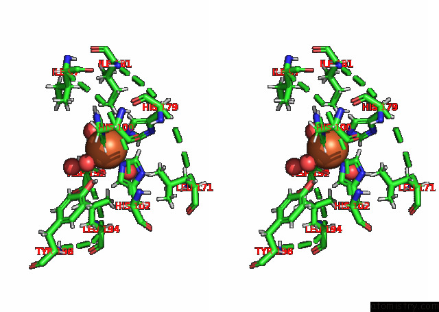
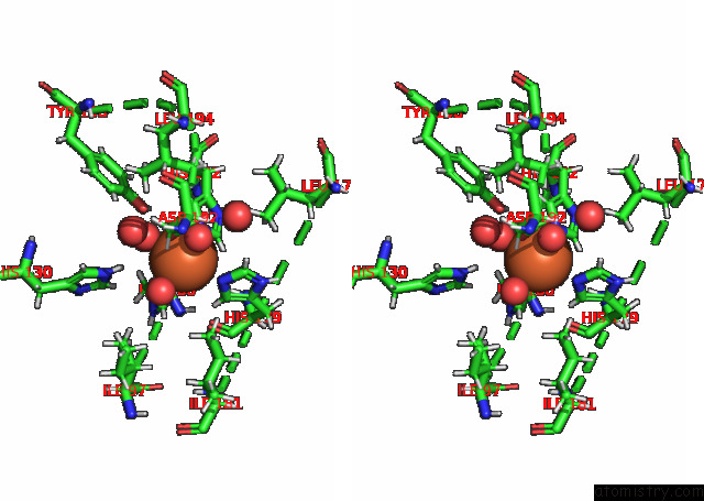
Iron binding site 4 out of 4 in 7lvz
Go back to
Iron binding site 4 out
of 4 in the Crystal Structure of Ado
Mono view
Stereo pair view
Mono view
Stereo pair view
A full contact list of Iron with other atoms in the Fe binding
site number 4 of Crystal Structure of Ado within 5.0Å range:
|
Reference:
R.L.Fernandez,
L.D.Elmendorf,
R.W.Smith,
C.A.Bingman,
B.G.Fox,
T.C.Brunold.
The Crystal Structure of Cysteamine Dioxygenase Reveals the Origin of the Large Substrate Scope of This Vital Mammalian Enzyme. Biochemistry V. 60 3728 2021.
ISSN: ISSN 0006-2960
PubMed: 34762398
DOI: 10.1021/ACS.BIOCHEM.1C00463
Page generated: Thu Aug 8 08:02:56 2024
ISSN: ISSN 0006-2960
PubMed: 34762398
DOI: 10.1021/ACS.BIOCHEM.1C00463
Last articles
Zn in 9JPJZn in 9JP7
Zn in 9JPK
Zn in 9JPL
Zn in 9GN6
Zn in 9GN7
Zn in 9GKU
Zn in 9GKW
Zn in 9GKX
Zn in 9GL0