Iron in PDB 7ttp: P450 (Oxya) From Kistamicin Biosynthesis, Mixed Heme Conformation
Protein crystallography data
The structure of P450 (Oxya) From Kistamicin Biosynthesis, Mixed Heme Conformation, PDB code: 7ttp
was solved by
A.Greule,
T.Izore,
M.J.Cryle,
with X-Ray Crystallography technique. A brief refinement statistics is given in the table below:
| Resolution Low / High (Å) | 35.66 / 1.80 |
| Space group | P 32 2 1 |
| Cell size a, b, c (Å), α, β, γ (°) | 69.806, 69.806, 132.456, 90, 90, 120 |
| R / Rfree (%) | 18.5 / 21.9 |
Iron Binding Sites:
The binding sites of Iron atom in the P450 (Oxya) From Kistamicin Biosynthesis, Mixed Heme Conformation
(pdb code 7ttp). This binding sites where shown within
5.0 Angstroms radius around Iron atom.
In total only one binding site of Iron was determined in the P450 (Oxya) From Kistamicin Biosynthesis, Mixed Heme Conformation, PDB code: 7ttp:
In total only one binding site of Iron was determined in the P450 (Oxya) From Kistamicin Biosynthesis, Mixed Heme Conformation, PDB code: 7ttp:
Iron binding site 1 out of 1 in 7ttp
Go back to
Iron binding site 1 out
of 1 in the P450 (Oxya) From Kistamicin Biosynthesis, Mixed Heme Conformation

Mono view
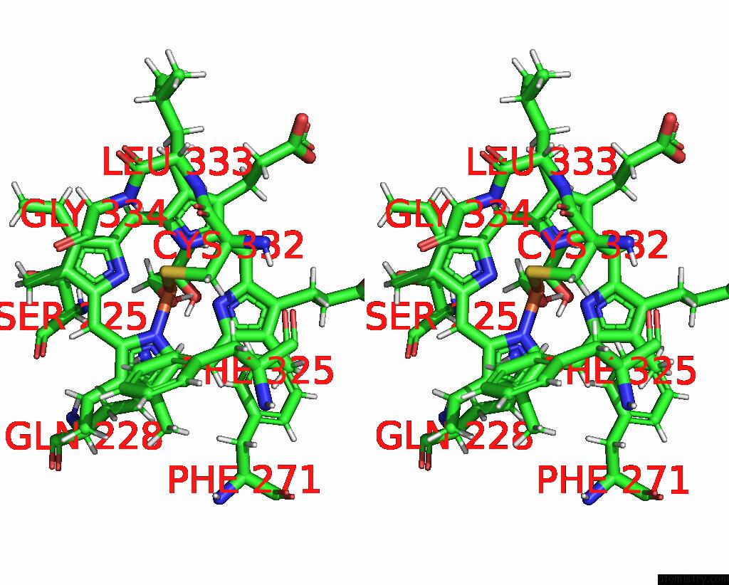
Stereo pair view
Mono view
Stereo pair view
A full contact list of Iron with other atoms in the Fe binding
site number 1 of P450 (Oxya) From Kistamicin Biosynthesis, Mixed Heme Conformation within 5.0Å range:
|
Reference:
A.Greule,
T.Izore,
D.Machell,
M.H.Hansen,
M.Schoppet,
J.J.De Voss,
L.K.Charkoudian,
R.B.Schittenhelm,
J.R.Harmer,
M.J.Cryle.
The Cytochrome P450 Oxya From the Kistamicin Biosynthesis Cyclization Cascade Is Highly Sensitive to Oxidative Damage. Front Chem V. 10 68240 2022.
ISSN: ESSN 2296-2646
PubMed: 35464232
DOI: 10.3389/FCHEM.2022.868240
Page generated: Fri Aug 9 02:40:50 2024
ISSN: ESSN 2296-2646
PubMed: 35464232
DOI: 10.3389/FCHEM.2022.868240
Last articles
Zn in 9J0NZn in 9J0O
Zn in 9J0P
Zn in 9FJX
Zn in 9EKB
Zn in 9C0F
Zn in 9CAH
Zn in 9CH0
Zn in 9CH3
Zn in 9CH1