Iron in PDB 8aq0: Crystal Structure of L-N-Carbamoylase From Sinorhizobium Meliloti Mutant L217G/F329C
Enzymatic activity of Crystal Structure of L-N-Carbamoylase From Sinorhizobium Meliloti Mutant L217G/F329C
All present enzymatic activity of Crystal Structure of L-N-Carbamoylase From Sinorhizobium Meliloti Mutant L217G/F329C:
3.5.1.87;
3.5.1.87;
Protein crystallography data
The structure of Crystal Structure of L-N-Carbamoylase From Sinorhizobium Meliloti Mutant L217G/F329C, PDB code: 8aq0
was solved by
H.J.Rozeboom,
C.Mayer,
with X-Ray Crystallography technique. A brief refinement statistics is given in the table below:
| Resolution Low / High (Å) | 99.23 / 2.30 |
| Space group | I 1 2 1 |
| Cell size a, b, c (Å), α, β, γ (°) | 132.569, 42.047, 137.211, 90, 94.78, 90 |
| R / Rfree (%) | 17.3 / 23.8 |
Other elements in 8aq0:
The structure of Crystal Structure of L-N-Carbamoylase From Sinorhizobium Meliloti Mutant L217G/F329C also contains other interesting chemical elements:
| Chlorine | (Cl) | 1 atom |
| Zinc | (Zn) | 4 atoms |
Iron Binding Sites:
The binding sites of Iron atom in the Crystal Structure of L-N-Carbamoylase From Sinorhizobium Meliloti Mutant L217G/F329C
(pdb code 8aq0). This binding sites where shown within
5.0 Angstroms radius around Iron atom.
In total 4 binding sites of Iron where determined in the Crystal Structure of L-N-Carbamoylase From Sinorhizobium Meliloti Mutant L217G/F329C, PDB code: 8aq0:
Jump to Iron binding site number: 1; 2; 3; 4;
In total 4 binding sites of Iron where determined in the Crystal Structure of L-N-Carbamoylase From Sinorhizobium Meliloti Mutant L217G/F329C, PDB code: 8aq0:
Jump to Iron binding site number: 1; 2; 3; 4;
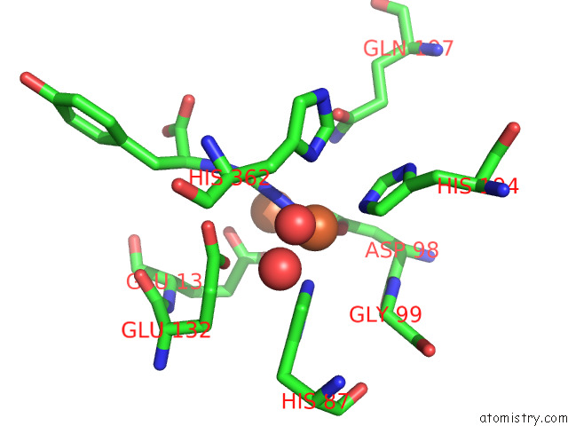
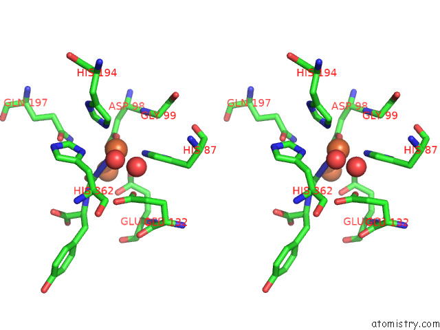
Iron binding site 1 out of 4 in 8aq0
Go back to
Iron binding site 1 out
of 4 in the Crystal Structure of L-N-Carbamoylase From Sinorhizobium Meliloti Mutant L217G/F329C
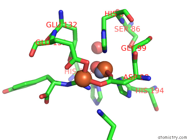
Mono view
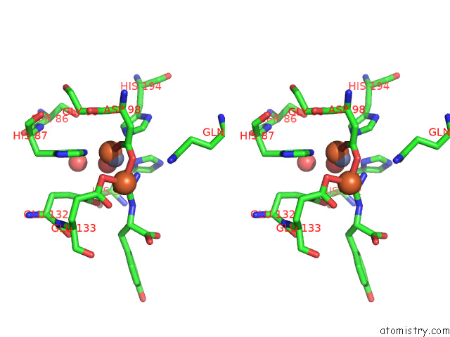
Stereo pair view
Mono view
Stereo pair view
A full contact list of Iron with other atoms in the Fe binding
site number 1 of Crystal Structure of L-N-Carbamoylase From Sinorhizobium Meliloti Mutant L217G/F329C within 5.0Å range:
|
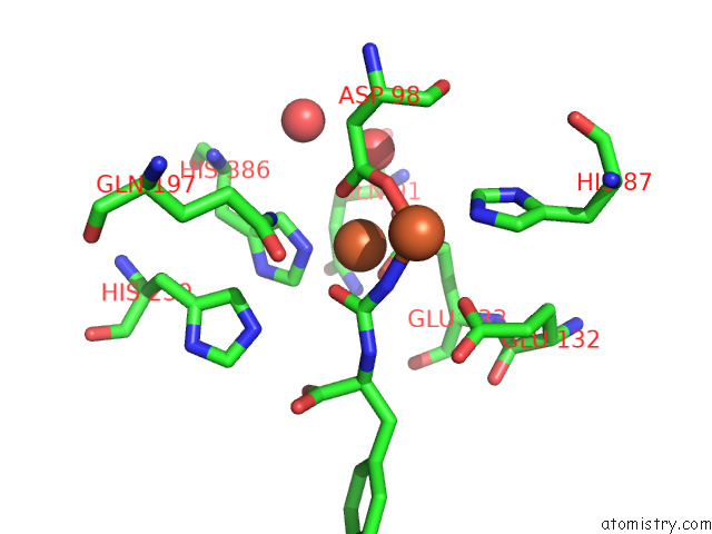
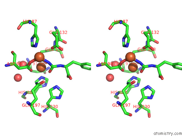
Iron binding site 2 out of 4 in 8aq0
Go back to
Iron binding site 2 out
of 4 in the Crystal Structure of L-N-Carbamoylase From Sinorhizobium Meliloti Mutant L217G/F329C
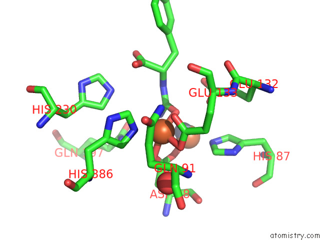
Mono view
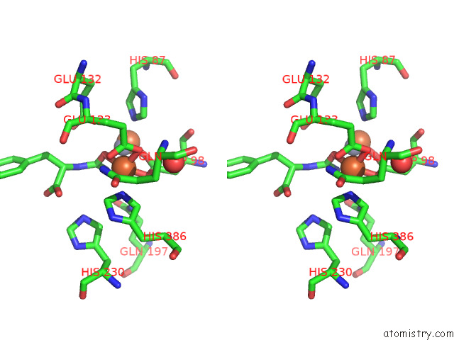
Stereo pair view
Mono view
Stereo pair view
A full contact list of Iron with other atoms in the Fe binding
site number 2 of Crystal Structure of L-N-Carbamoylase From Sinorhizobium Meliloti Mutant L217G/F329C within 5.0Å range:
|
Iron binding site 3 out of 4 in 8aq0
Go back to
Iron binding site 3 out
of 4 in the Crystal Structure of L-N-Carbamoylase From Sinorhizobium Meliloti Mutant L217G/F329C
Mono view
Stereo pair view
Mono view
Stereo pair view
A full contact list of Iron with other atoms in the Fe binding
site number 3 of Crystal Structure of L-N-Carbamoylase From Sinorhizobium Meliloti Mutant L217G/F329C within 5.0Å range:
|
Iron binding site 4 out of 4 in 8aq0
Go back to
Iron binding site 4 out
of 4 in the Crystal Structure of L-N-Carbamoylase From Sinorhizobium Meliloti Mutant L217G/F329C
Mono view
Stereo pair view
Mono view
Stereo pair view
A full contact list of Iron with other atoms in the Fe binding
site number 4 of Crystal Structure of L-N-Carbamoylase From Sinorhizobium Meliloti Mutant L217G/F329C within 5.0Å range:
|
Reference:
R.Rubini,
S.C.Jansen,
H.Beekhuis,
H.J.Rozeboom,
C.Mayer.
Selecting Better Biocatalysts By Complementing Recoded Bacteria. Angew.Chem.Int.Ed.Engl. 13942 2022.
ISSN: ESSN 1521-3773
PubMed: 36342942
DOI: 10.1002/ANIE.202213942
Page generated: Fri Aug 9 18:36:42 2024
ISSN: ESSN 1521-3773
PubMed: 36342942
DOI: 10.1002/ANIE.202213942
Last articles
Zn in 9J0NZn in 9J0O
Zn in 9J0P
Zn in 9FJX
Zn in 9EKB
Zn in 9C0F
Zn in 9CAH
Zn in 9CH0
Zn in 9CH3
Zn in 9CH1