Iron in PDB 8k6y: Serial Femtosecond Crystallography Structure of Photo Dissociated Co From BA3- Type Cytochrome C Oxidase Determined By Extrapolation Method
Enzymatic activity of Serial Femtosecond Crystallography Structure of Photo Dissociated Co From BA3- Type Cytochrome C Oxidase Determined By Extrapolation Method
All present enzymatic activity of Serial Femtosecond Crystallography Structure of Photo Dissociated Co From BA3- Type Cytochrome C Oxidase Determined By Extrapolation Method:
7.1.1.9;
7.1.1.9;
Protein crystallography data
The structure of Serial Femtosecond Crystallography Structure of Photo Dissociated Co From BA3- Type Cytochrome C Oxidase Determined By Extrapolation Method, PDB code: 8k6y
was solved by
C.Safari,
S.Ghosh,
R.Andersson,
J.Johannesson,
A.V.Donoso,
D.Zoric,
E.Sandelin,
S.Iwata,
R.Neutze,
G.Branden,
with X-Ray Crystallography technique. A brief refinement statistics is given in the table below:
| Resolution Low / High (Å) | 37.15 / 2.00 |
| Space group | C 1 2 1 |
| Cell size a, b, c (Å), α, β, γ (°) | 145.85, 100.32, 96.62, 90, 126.76, 90 |
| R / Rfree (%) | 39.6 / 43.5 |
Other elements in 8k6y:
The structure of Serial Femtosecond Crystallography Structure of Photo Dissociated Co From BA3- Type Cytochrome C Oxidase Determined By Extrapolation Method also contains other interesting chemical elements:
| Copper | (Cu) | 3 atoms |
Iron Binding Sites:
The binding sites of Iron atom in the Serial Femtosecond Crystallography Structure of Photo Dissociated Co From BA3- Type Cytochrome C Oxidase Determined By Extrapolation Method
(pdb code 8k6y). This binding sites where shown within
5.0 Angstroms radius around Iron atom.
In total 2 binding sites of Iron where determined in the Serial Femtosecond Crystallography Structure of Photo Dissociated Co From BA3- Type Cytochrome C Oxidase Determined By Extrapolation Method, PDB code: 8k6y:
Jump to Iron binding site number: 1; 2;
In total 2 binding sites of Iron where determined in the Serial Femtosecond Crystallography Structure of Photo Dissociated Co From BA3- Type Cytochrome C Oxidase Determined By Extrapolation Method, PDB code: 8k6y:
Jump to Iron binding site number: 1; 2;
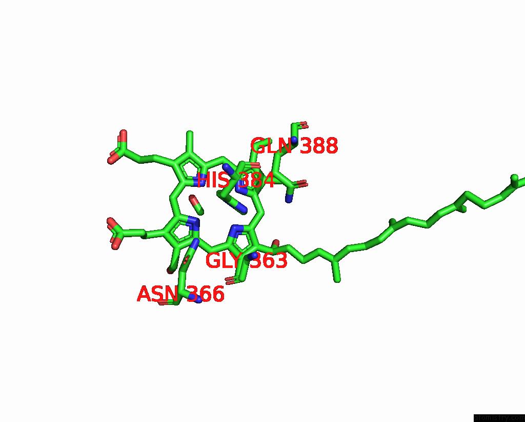
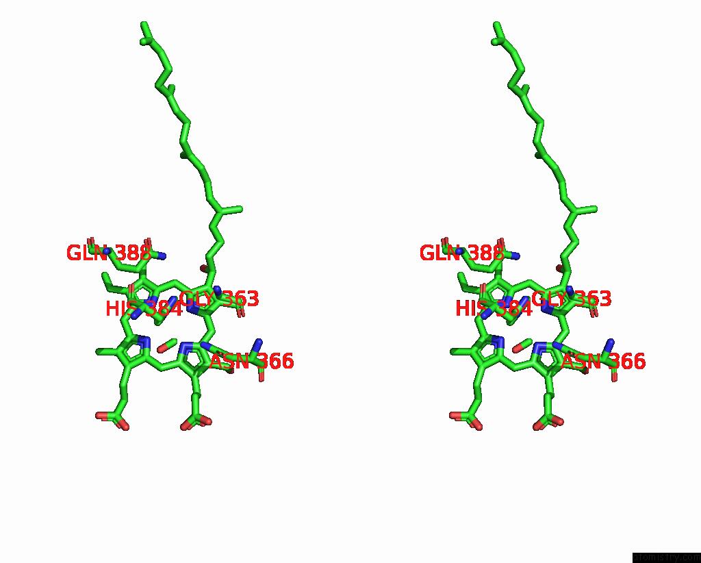
Iron binding site 1 out of 2 in 8k6y
Go back to
Iron binding site 1 out
of 2 in the Serial Femtosecond Crystallography Structure of Photo Dissociated Co From BA3- Type Cytochrome C Oxidase Determined By Extrapolation Method

Mono view

Stereo pair view
Mono view
Stereo pair view
A full contact list of Iron with other atoms in the Fe binding
site number 1 of Serial Femtosecond Crystallography Structure of Photo Dissociated Co From BA3- Type Cytochrome C Oxidase Determined By Extrapolation Method within 5.0Å range:
|
Iron binding site 2 out of 2 in 8k6y
Go back to
Iron binding site 2 out
of 2 in the Serial Femtosecond Crystallography Structure of Photo Dissociated Co From BA3- Type Cytochrome C Oxidase Determined By Extrapolation Method
Mono view
Stereo pair view
Mono view
Stereo pair view
A full contact list of Iron with other atoms in the Fe binding
site number 2 of Serial Femtosecond Crystallography Structure of Photo Dissociated Co From BA3- Type Cytochrome C Oxidase Determined By Extrapolation Method within 5.0Å range:
|
Reference:
C.Safari,
S.Ghosh,
R.Andersson,
J.Johannesson,
A.V.Donoso,
D.Zoric,
E.Sandelin,
S.Iwata,
R.Neutze,
G.Branden.
Serial Femtosecond Crystallography Structure of Photo Dissociated Co From BA3- Type Cytochrome C Oxidase Determined By Extrapolation Method To Be Published.
Page generated: Sat Aug 10 07:27:16 2024
Last articles
Zn in 9J0NZn in 9J0O
Zn in 9J0P
Zn in 9FJX
Zn in 9EKB
Zn in 9C0F
Zn in 9CAH
Zn in 9CH0
Zn in 9CH3
Zn in 9CH1