Iron in PDB 8kht: The Structure of RV0097 with Substrate
Protein crystallography data
The structure of The Structure of RV0097 with Substrate, PDB code: 8kht
was solved by
J.Chen,
J.Zhou,
with X-Ray Crystallography technique. A brief refinement statistics is given in the table below:
| Resolution Low / High (Å) | 28.37 / 2.05 |
| Space group | P 1 21 1 |
| Cell size a, b, c (Å), α, β, γ (°) | 56.74, 89.07, 63.51, 90, 90.03, 90 |
| R / Rfree (%) | 20.7 / 26.6 |
Iron Binding Sites:
The binding sites of Iron atom in the The Structure of RV0097 with Substrate
(pdb code 8kht). This binding sites where shown within
5.0 Angstroms radius around Iron atom.
In total 2 binding sites of Iron where determined in the The Structure of RV0097 with Substrate, PDB code: 8kht:
Jump to Iron binding site number: 1; 2;
In total 2 binding sites of Iron where determined in the The Structure of RV0097 with Substrate, PDB code: 8kht:
Jump to Iron binding site number: 1; 2;
Iron binding site 1 out of 2 in 8kht
Go back to
Iron binding site 1 out
of 2 in the The Structure of RV0097 with Substrate
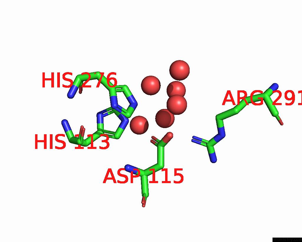
Mono view
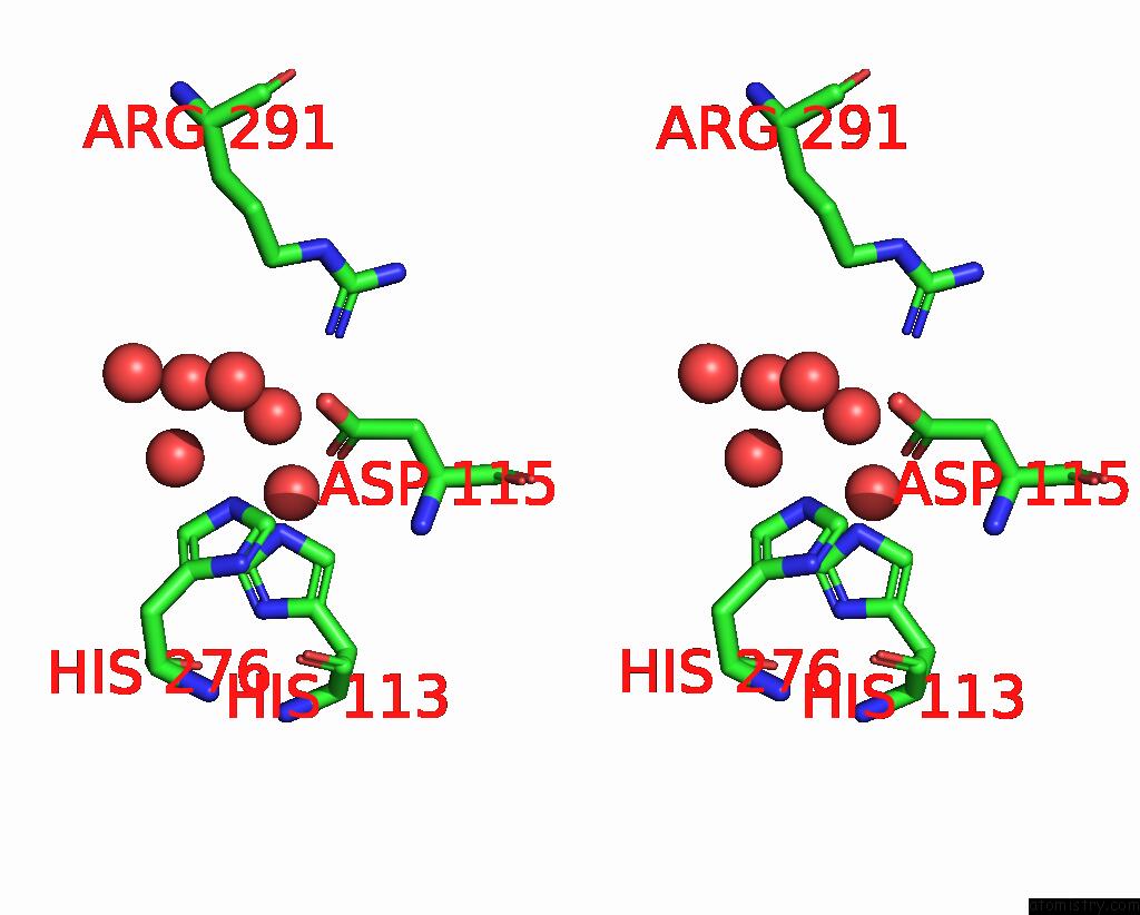
Stereo pair view
Mono view
Stereo pair view
A full contact list of Iron with other atoms in the Fe binding
site number 1 of The Structure of RV0097 with Substrate within 5.0Å range:
|
Iron binding site 2 out of 2 in 8kht
Go back to
Iron binding site 2 out
of 2 in the The Structure of RV0097 with Substrate
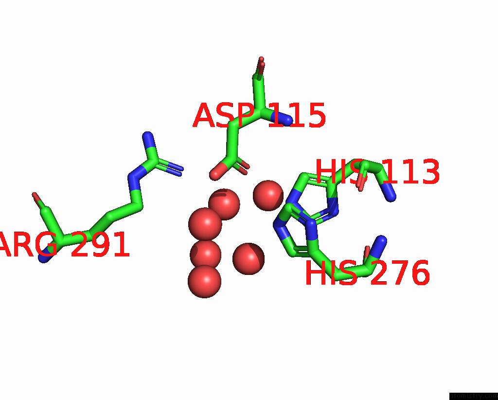
Mono view
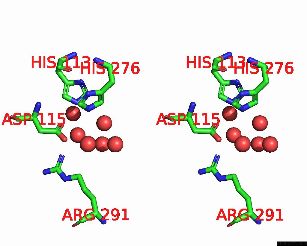
Stereo pair view
Mono view
Stereo pair view
A full contact list of Iron with other atoms in the Fe binding
site number 2 of The Structure of RV0097 with Substrate within 5.0Å range:
|
Reference:
T.Y.Chen,
J.Chen,
M.W.Ruszczycky,
D.Hilovsky,
T.Hostetler,
X.Liu,
J.Zhou,
W.Chang.
Variation in Biosynthesis and Metal-Binding Properties of Isonitrile-Containing Peptides Produced By Mycobacteria Versus Streptomyces. Acs Catalysis V. 14 4975 2024.
ISSN: ESSN 2155-5435
DOI: 10.1021/ACSCATAL.4C00645
Page generated: Sat Aug 10 07:38:43 2024
ISSN: ESSN 2155-5435
DOI: 10.1021/ACSCATAL.4C00645
Last articles
Zn in 9J0NZn in 9J0O
Zn in 9J0P
Zn in 9FJX
Zn in 9EKB
Zn in 9C0F
Zn in 9CAH
Zn in 9CH0
Zn in 9CH3
Zn in 9CH1