Iron in PDB 8ki1: Crystal Structure of the Holo Form of the Hemophore Hasa From Pseudomonas Protegens Pf-5
Protein crystallography data
The structure of Crystal Structure of the Holo Form of the Hemophore Hasa From Pseudomonas Protegens Pf-5, PDB code: 8ki1
was solved by
Y.Shisaka,
H.Inaba,
H.Sugimoto,
O.Shoji,
with X-Ray Crystallography technique. A brief refinement statistics is given in the table below:
| Resolution Low / High (Å) | 20.00 / 1.90 |
| Space group | P 65 |
| Cell size a, b, c (Å), α, β, γ (°) | 99.597, 99.597, 90.473, 90, 90, 120 |
| R / Rfree (%) | 15.8 / 19.3 |
Iron Binding Sites:
The binding sites of Iron atom in the Crystal Structure of the Holo Form of the Hemophore Hasa From Pseudomonas Protegens Pf-5
(pdb code 8ki1). This binding sites where shown within
5.0 Angstroms radius around Iron atom.
In total 2 binding sites of Iron where determined in the Crystal Structure of the Holo Form of the Hemophore Hasa From Pseudomonas Protegens Pf-5, PDB code: 8ki1:
Jump to Iron binding site number: 1; 2;
In total 2 binding sites of Iron where determined in the Crystal Structure of the Holo Form of the Hemophore Hasa From Pseudomonas Protegens Pf-5, PDB code: 8ki1:
Jump to Iron binding site number: 1; 2;
Iron binding site 1 out of 2 in 8ki1
Go back to
Iron binding site 1 out
of 2 in the Crystal Structure of the Holo Form of the Hemophore Hasa From Pseudomonas Protegens Pf-5
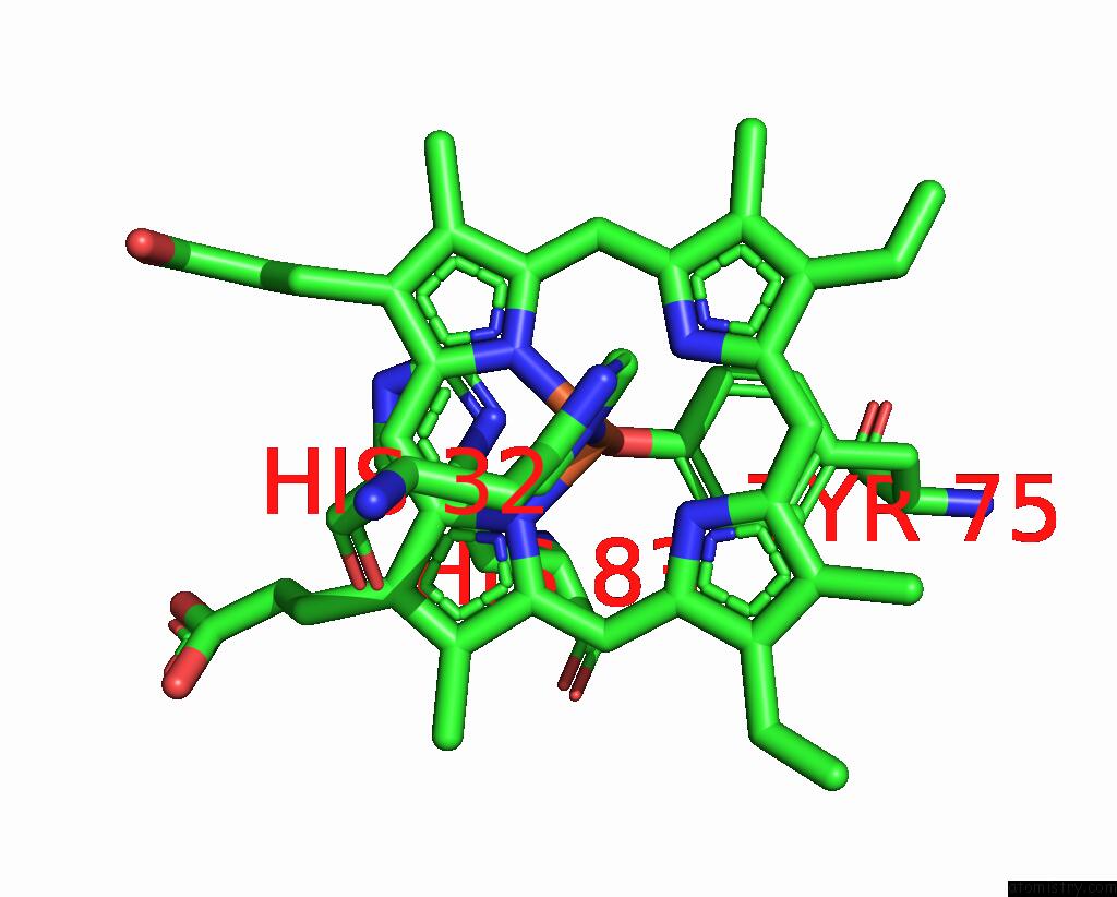
Mono view
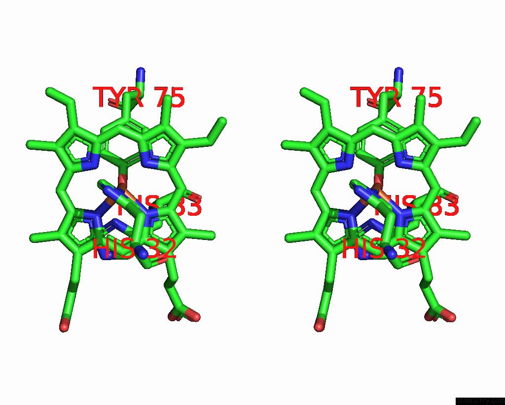
Stereo pair view
Mono view
Stereo pair view
A full contact list of Iron with other atoms in the Fe binding
site number 1 of Crystal Structure of the Holo Form of the Hemophore Hasa From Pseudomonas Protegens Pf-5 within 5.0Å range:
|
Iron binding site 2 out of 2 in 8ki1
Go back to
Iron binding site 2 out
of 2 in the Crystal Structure of the Holo Form of the Hemophore Hasa From Pseudomonas Protegens Pf-5
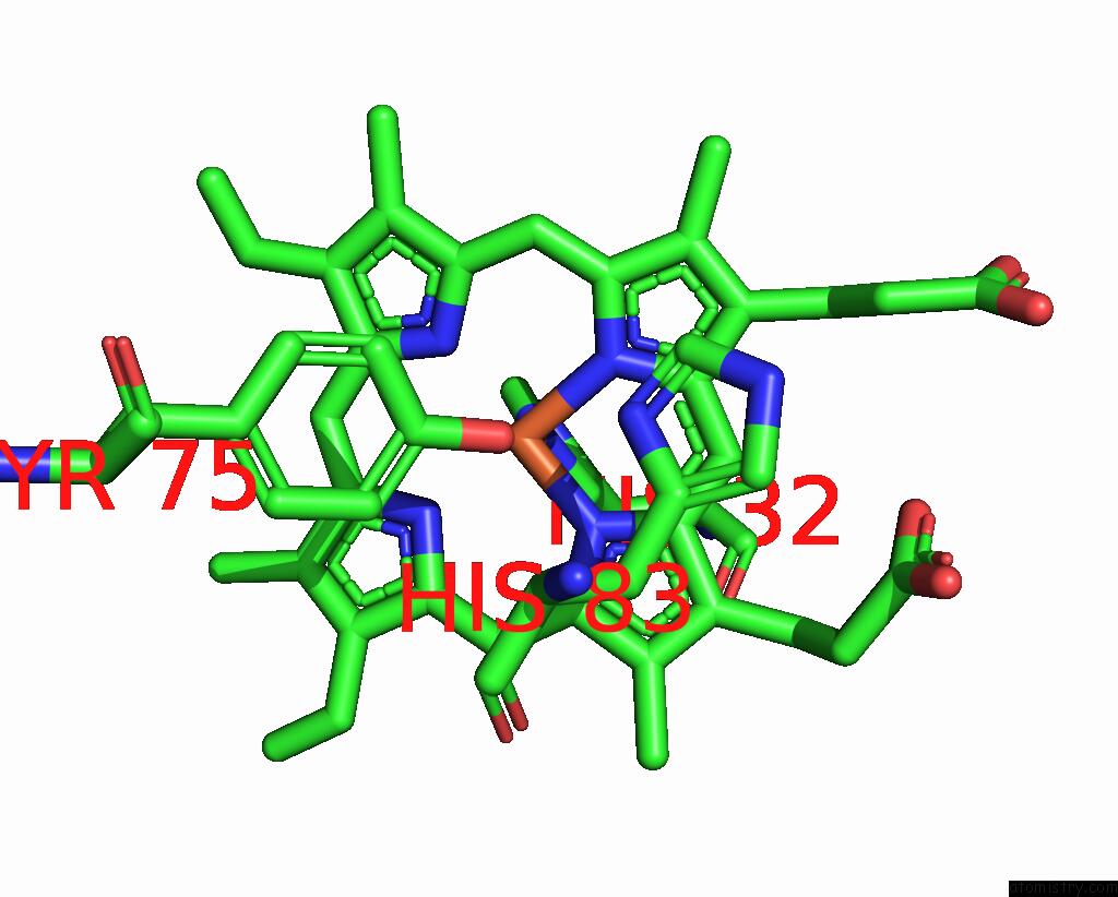
Mono view
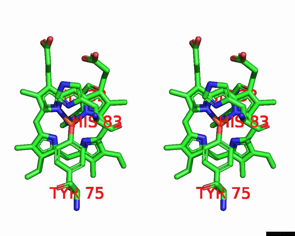
Stereo pair view
Mono view
Stereo pair view
A full contact list of Iron with other atoms in the Fe binding
site number 2 of Crystal Structure of the Holo Form of the Hemophore Hasa From Pseudomonas Protegens Pf-5 within 5.0Å range:
|
Reference:
H.Inaba,
Y.Shisaka,
S.Ariyasu,
E.Sakakibara,
G.Ueda,
Y.Aiba,
N.Shimizu,
H.Sugimoto,
O.Shoji.
Heme-Substituted Protein Assembly Bridged By Synthetic Porphyrin: Achieving Controlled Configuration While Maintaining Rotational Freedom Rsc Adv V. 14 8829 2024.
ISSN: ESSN 2046-2069
DOI: 10.1039/D4RA01042F
Page generated: Sat Aug 10 07:39:10 2024
ISSN: ESSN 2046-2069
DOI: 10.1039/D4RA01042F
Last articles
Zn in 9J0NZn in 9J0O
Zn in 9J0P
Zn in 9FJX
Zn in 9EKB
Zn in 9C0F
Zn in 9CAH
Zn in 9CH0
Zn in 9CH3
Zn in 9CH1