Iron in PDB 8s1j: Crystal Structure of T-Anethole Oxygenase From Stenotrophomonas Maltophilia
Protein crystallography data
The structure of Crystal Structure of T-Anethole Oxygenase From Stenotrophomonas Maltophilia, PDB code: 8s1j
was solved by
H.J.Rozeboom,
M.W.Fraaije,
with X-Ray Crystallography technique. A brief refinement statistics is given in the table below:
| Resolution Low / High (Å) | 58.80 / 1.83 |
| Space group | P 1 21 1 |
| Cell size a, b, c (Å), α, β, γ (°) | 51.254, 96.135, 78.311, 90, 108.66, 90 |
| R / Rfree (%) | 19.2 / 22.8 |
Other elements in 8s1j:
The structure of Crystal Structure of T-Anethole Oxygenase From Stenotrophomonas Maltophilia also contains other interesting chemical elements:
| Chlorine | (Cl) | 3 atoms |
Iron Binding Sites:
The binding sites of Iron atom in the Crystal Structure of T-Anethole Oxygenase From Stenotrophomonas Maltophilia
(pdb code 8s1j). This binding sites where shown within
5.0 Angstroms radius around Iron atom.
In total 2 binding sites of Iron where determined in the Crystal Structure of T-Anethole Oxygenase From Stenotrophomonas Maltophilia, PDB code: 8s1j:
Jump to Iron binding site number: 1; 2;
In total 2 binding sites of Iron where determined in the Crystal Structure of T-Anethole Oxygenase From Stenotrophomonas Maltophilia, PDB code: 8s1j:
Jump to Iron binding site number: 1; 2;
Iron binding site 1 out of 2 in 8s1j
Go back to
Iron binding site 1 out
of 2 in the Crystal Structure of T-Anethole Oxygenase From Stenotrophomonas Maltophilia
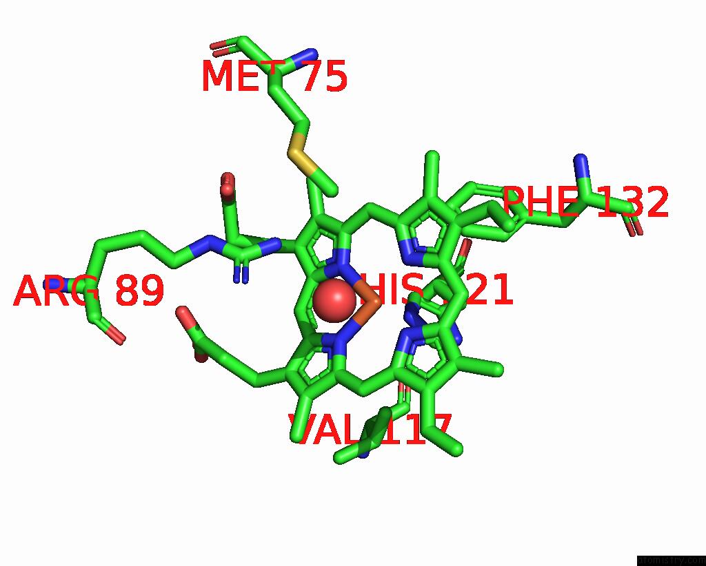
Mono view
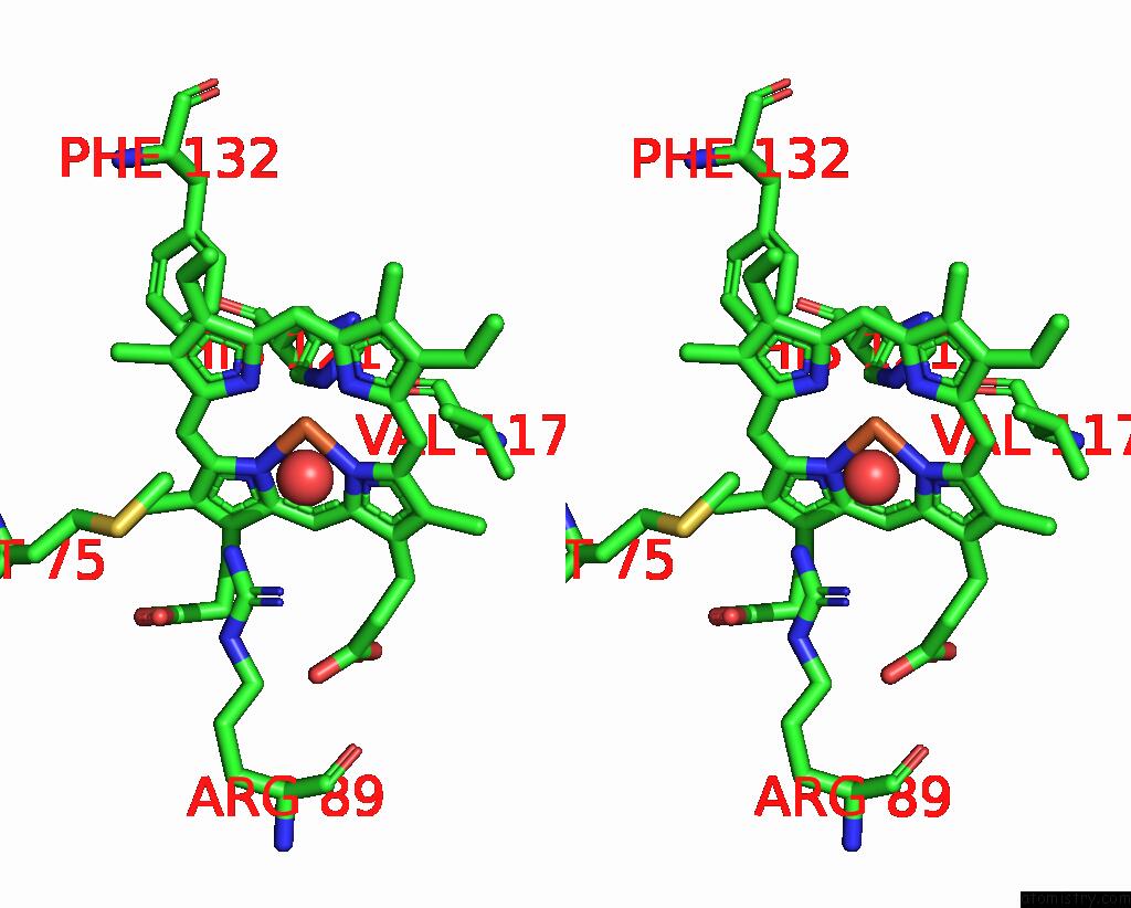
Stereo pair view
Mono view
Stereo pair view
A full contact list of Iron with other atoms in the Fe binding
site number 1 of Crystal Structure of T-Anethole Oxygenase From Stenotrophomonas Maltophilia within 5.0Å range:
|
Iron binding site 2 out of 2 in 8s1j
Go back to
Iron binding site 2 out
of 2 in the Crystal Structure of T-Anethole Oxygenase From Stenotrophomonas Maltophilia
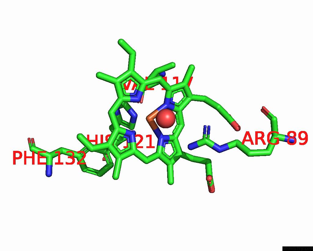
Mono view
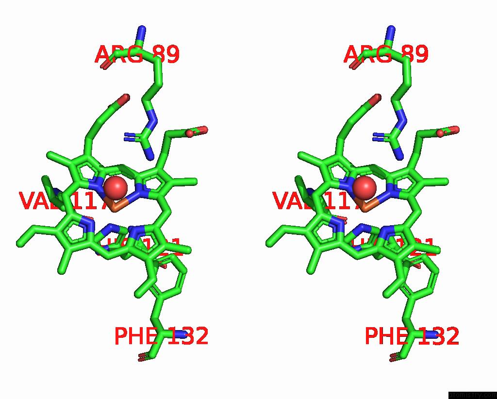
Stereo pair view
Mono view
Stereo pair view
A full contact list of Iron with other atoms in the Fe binding
site number 2 of Crystal Structure of T-Anethole Oxygenase From Stenotrophomonas Maltophilia within 5.0Å range:
|
Reference:
N.N.Purwani,
H.J.Rozeboom,
V.P.Willers,
H.J.Wijma,
M.W.Fraaije.
Discovery of A New Class of Bacterial Heme-Containing Cc Cleaving Oxygenases. N Biotechnol V. 83 82 2024.
ISSN: ESSN 1876-4347
PubMed: 39053683
DOI: 10.1016/J.NBT.2024.07.002
Page generated: Sat Sep 28 21:45:52 2024
ISSN: ESSN 1876-4347
PubMed: 39053683
DOI: 10.1016/J.NBT.2024.07.002
Last articles
Zn in 9MJ5Zn in 9HNW
Zn in 9G0L
Zn in 9FNE
Zn in 9DZN
Zn in 9E0I
Zn in 9D32
Zn in 9DAK
Zn in 8ZXC
Zn in 8ZUF