Iron in PDB 8sbi: Crystal Structure of Human Sterol 14 Alpha-Demethylase (CYP51) in the Ligand-Free State
Enzymatic activity of Crystal Structure of Human Sterol 14 Alpha-Demethylase (CYP51) in the Ligand-Free State
All present enzymatic activity of Crystal Structure of Human Sterol 14 Alpha-Demethylase (CYP51) in the Ligand-Free State:
1.14.14.154;
1.14.14.154;
Protein crystallography data
The structure of Crystal Structure of Human Sterol 14 Alpha-Demethylase (CYP51) in the Ligand-Free State, PDB code: 8sbi
was solved by
T.Y.Hargrove,
Z.Wawrzak,
G.I.Lepesheva,
with X-Ray Crystallography technique. A brief refinement statistics is given in the table below:
| Resolution Low / High (Å) | 49.01 / 2.73 |
| Space group | P 21 21 2 |
| Cell size a, b, c (Å), α, β, γ (°) | 143.81, 55.64, 103.07, 90, 90, 90 |
| R / Rfree (%) | 23 / 24.6 |
Iron Binding Sites:
The binding sites of Iron atom in the Crystal Structure of Human Sterol 14 Alpha-Demethylase (CYP51) in the Ligand-Free State
(pdb code 8sbi). This binding sites where shown within
5.0 Angstroms radius around Iron atom.
In total 2 binding sites of Iron where determined in the Crystal Structure of Human Sterol 14 Alpha-Demethylase (CYP51) in the Ligand-Free State, PDB code: 8sbi:
Jump to Iron binding site number: 1; 2;
In total 2 binding sites of Iron where determined in the Crystal Structure of Human Sterol 14 Alpha-Demethylase (CYP51) in the Ligand-Free State, PDB code: 8sbi:
Jump to Iron binding site number: 1; 2;
Iron binding site 1 out of 2 in 8sbi
Go back to
Iron binding site 1 out
of 2 in the Crystal Structure of Human Sterol 14 Alpha-Demethylase (CYP51) in the Ligand-Free State
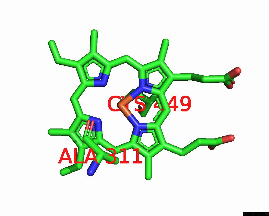
Mono view
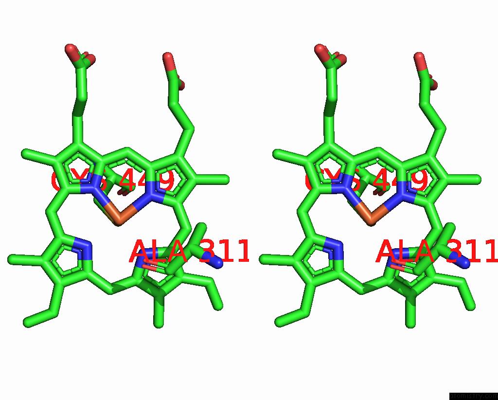
Stereo pair view
Mono view
Stereo pair view
A full contact list of Iron with other atoms in the Fe binding
site number 1 of Crystal Structure of Human Sterol 14 Alpha-Demethylase (CYP51) in the Ligand-Free State within 5.0Å range:
|
Iron binding site 2 out of 2 in 8sbi
Go back to
Iron binding site 2 out
of 2 in the Crystal Structure of Human Sterol 14 Alpha-Demethylase (CYP51) in the Ligand-Free State

Mono view
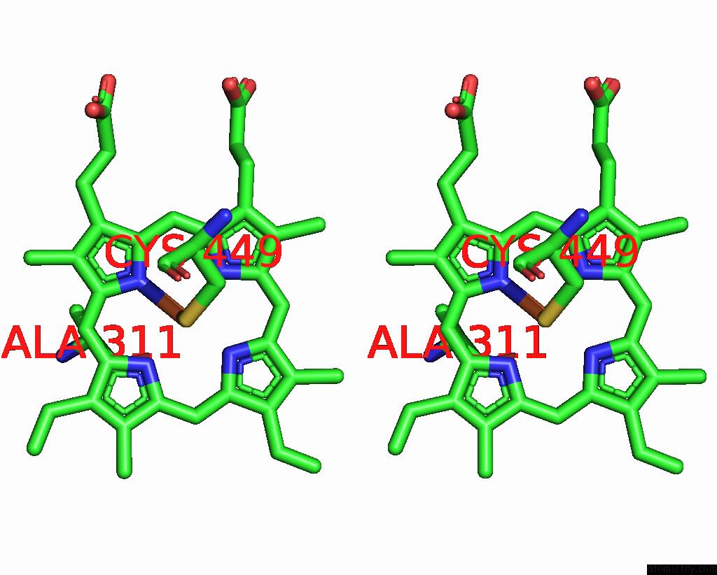
Stereo pair view
Mono view
Stereo pair view
A full contact list of Iron with other atoms in the Fe binding
site number 2 of Crystal Structure of Human Sterol 14 Alpha-Demethylase (CYP51) in the Ligand-Free State within 5.0Å range:
|
Reference:
T.Y.Hargrove,
Z.Wawrzak,
G.I.Lepesheva.
Structural Dynamics of Sterol 14 Alpha Demethylases Upon Catalysis To Be Published.
Page generated: Sat Aug 10 17:46:11 2024
Last articles
Zn in 9IRQZn in 9IYX
Zn in 9J8P
Zn in 9IUU
Zn in 9GBF
Zn in 9G2V
Zn in 9G2L
Zn in 9G2X
Zn in 9G2Z
Zn in 9G2K