Iron in PDB 8taw: The Crystal Structure of T252E CYP199A4 Bound to 4-(Pyridin-2-Yl) Benzoic Acid
Protein crystallography data
The structure of The Crystal Structure of T252E CYP199A4 Bound to 4-(Pyridin-2-Yl) Benzoic Acid, PDB code: 8taw
was solved by
M.N.Podgorski,
S.G.Bell,
with X-Ray Crystallography technique. A brief refinement statistics is given in the table below:
| Resolution Low / High (Å) | 44.13 / 1.73 |
| Space group | P 1 21 1 |
| Cell size a, b, c (Å), α, β, γ (°) | 44.17, 51.52, 79.23, 90, 92.3, 90 |
| R / Rfree (%) | 15.2 / 18.5 |
Other elements in 8taw:
The structure of The Crystal Structure of T252E CYP199A4 Bound to 4-(Pyridin-2-Yl) Benzoic Acid also contains other interesting chemical elements:
| Chlorine | (Cl) | 1 atom |
Iron Binding Sites:
The binding sites of Iron atom in the The Crystal Structure of T252E CYP199A4 Bound to 4-(Pyridin-2-Yl) Benzoic Acid
(pdb code 8taw). This binding sites where shown within
5.0 Angstroms radius around Iron atom.
In total only one binding site of Iron was determined in the The Crystal Structure of T252E CYP199A4 Bound to 4-(Pyridin-2-Yl) Benzoic Acid, PDB code: 8taw:
In total only one binding site of Iron was determined in the The Crystal Structure of T252E CYP199A4 Bound to 4-(Pyridin-2-Yl) Benzoic Acid, PDB code: 8taw:
Iron binding site 1 out of 1 in 8taw
Go back to
Iron binding site 1 out
of 1 in the The Crystal Structure of T252E CYP199A4 Bound to 4-(Pyridin-2-Yl) Benzoic Acid
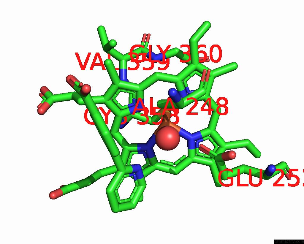
Mono view
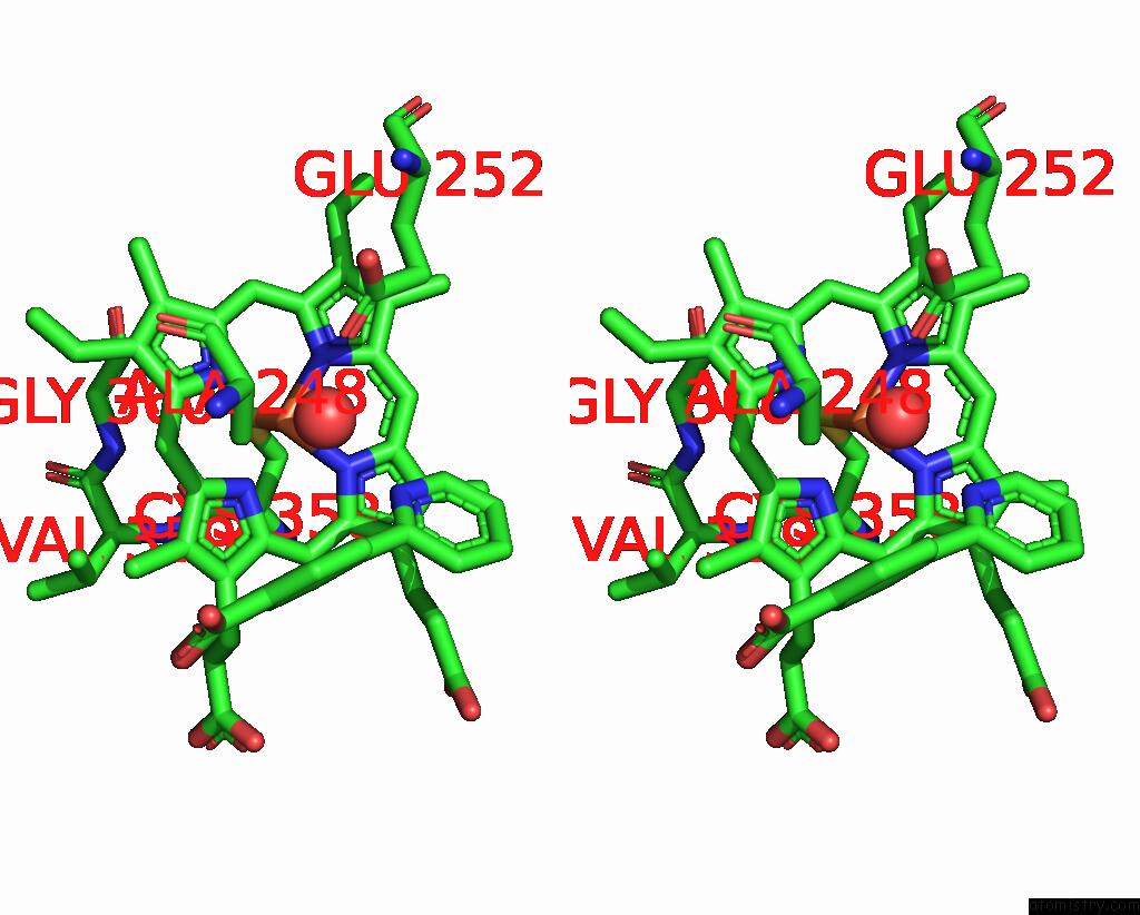
Stereo pair view
Mono view
Stereo pair view
A full contact list of Iron with other atoms in the Fe binding
site number 1 of The Crystal Structure of T252E CYP199A4 Bound to 4-(Pyridin-2-Yl) Benzoic Acid within 5.0Å range:
|
Reference:
M.N.Podgorski,
J.H.Z.Lee,
J.S.Harbort,
G.T.H.Nguyen,
D.Z.Doherty,
W.A.Donald,
J.R.Harmer,
J.B.Bruning,
S.G.Bell.
Characterisation of the Heme Aqua-Ligand Coordination Environment in An Engineered Peroxygenase Cytochrome P450 Variant. J.Inorg.Biochem. V. 249 12391 2023.
ISSN: ISSN 0162-0134
PubMed: 37837941
DOI: 10.1016/J.JINORGBIO.2023.112391
Page generated: Sat Aug 10 18:18:11 2024
ISSN: ISSN 0162-0134
PubMed: 37837941
DOI: 10.1016/J.JINORGBIO.2023.112391
Last articles
Zn in 9IRQZn in 9IYX
Zn in 9J8P
Zn in 9IUU
Zn in 9GBF
Zn in 9G2V
Zn in 9G2L
Zn in 9G2X
Zn in 9G2Z
Zn in 9G2K