Iron in PDB 8tdq: Sfx-Xfel Structure of CYP121 Cocrystallized with Substrate Cyy
Enzymatic activity of Sfx-Xfel Structure of CYP121 Cocrystallized with Substrate Cyy
All present enzymatic activity of Sfx-Xfel Structure of CYP121 Cocrystallized with Substrate Cyy:
1.14.19.70;
1.14.19.70;
Protein crystallography data
The structure of Sfx-Xfel Structure of CYP121 Cocrystallized with Substrate Cyy, PDB code: 8tdq
was solved by
R.C.Nguyen,
M.Dasgupta,
A.Bhowmick,
J.F.Kern,
A.Liu,
with X-Ray Crystallography technique. A brief refinement statistics is given in the table below:
| Resolution Low / High (Å) | 31.77 / 1.65 |
| Space group | P 65 2 2 |
| Cell size a, b, c (Å), α, β, γ (°) | 78.616, 78.616, 265.275, 90, 90, 120 |
| R / Rfree (%) | 17.1 / 20.1 |
Iron Binding Sites:
The binding sites of Iron atom in the Sfx-Xfel Structure of CYP121 Cocrystallized with Substrate Cyy
(pdb code 8tdq). This binding sites where shown within
5.0 Angstroms radius around Iron atom.
In total only one binding site of Iron was determined in the Sfx-Xfel Structure of CYP121 Cocrystallized with Substrate Cyy, PDB code: 8tdq:
In total only one binding site of Iron was determined in the Sfx-Xfel Structure of CYP121 Cocrystallized with Substrate Cyy, PDB code: 8tdq:
Iron binding site 1 out of 1 in 8tdq
Go back to
Iron binding site 1 out
of 1 in the Sfx-Xfel Structure of CYP121 Cocrystallized with Substrate Cyy
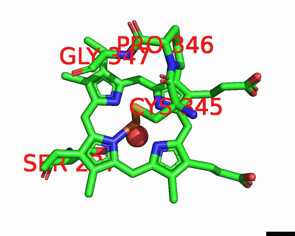
Mono view
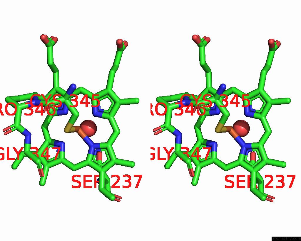
Stereo pair view
Mono view
Stereo pair view
A full contact list of Iron with other atoms in the Fe binding
site number 1 of Sfx-Xfel Structure of CYP121 Cocrystallized with Substrate Cyy within 5.0Å range:
|
Reference:
R.C.Nguyen,
I.Davis,
M.Dasgupta,
Y.Wang,
P.S.Simon,
A.Butryn,
H.Makita,
I.Bogacz,
K.Dornevil,
P.Aller,
A.Bhowmick,
R.Chatterjee,
I.S.Kim,
T.Zhou,
D.Mendez,
D.W.Paley,
F.Fuller,
R.Alonso Mori,
A.Batyuk,
N.K.Sauter,
A.S.Brewster,
A.M.Orville,
V.K.Yachandra,
J.Yano,
J.F.Kern,
A.Liu.
In Situ Structural Observation of A Substrate- and Peroxide-Bound High-Spin Ferric-Hydroperoxo Intermediate in the P450 Enzyme CYP121. J.Am.Chem.Soc. 2023.
ISSN: ESSN 1520-5126
PubMed: 37939223
DOI: 10.1021/JACS.3C04991
Page generated: Sat Aug 10 18:18:16 2024
ISSN: ESSN 1520-5126
PubMed: 37939223
DOI: 10.1021/JACS.3C04991
Last articles
Zn in 9J0NZn in 9J0O
Zn in 9J0P
Zn in 9FJX
Zn in 9EKB
Zn in 9C0F
Zn in 9CAH
Zn in 9CH0
Zn in 9CH3
Zn in 9CH1