Iron in PDB 8ugz: Crystal Structure of Shewanella Benthica Group 1 Truncated Hemoglobin C51S C71S Variant
Protein crystallography data
The structure of Crystal Structure of Shewanella Benthica Group 1 Truncated Hemoglobin C51S C71S Variant, PDB code: 8ugz
was solved by
T.D.Schultz,
J.E.Martinez,
M.A.Siegler,
J.L.Schlessman,
J.T.J.Lecomte,
with X-Ray Crystallography technique. A brief refinement statistics is given in the table below:
| Resolution Low / High (Å) | 23.29 / 1.80 |
| Space group | P 21 21 21 |
| Cell size a, b, c (Å), α, β, γ (°) | 27.486, 105.058, 151.118, 90, 90, 90 |
| R / Rfree (%) | 17.6 / 21 |
Iron Binding Sites:
The binding sites of Iron atom in the Crystal Structure of Shewanella Benthica Group 1 Truncated Hemoglobin C51S C71S Variant
(pdb code 8ugz). This binding sites where shown within
5.0 Angstroms radius around Iron atom.
In total 4 binding sites of Iron where determined in the Crystal Structure of Shewanella Benthica Group 1 Truncated Hemoglobin C51S C71S Variant, PDB code: 8ugz:
Jump to Iron binding site number: 1; 2; 3; 4;
In total 4 binding sites of Iron where determined in the Crystal Structure of Shewanella Benthica Group 1 Truncated Hemoglobin C51S C71S Variant, PDB code: 8ugz:
Jump to Iron binding site number: 1; 2; 3; 4;
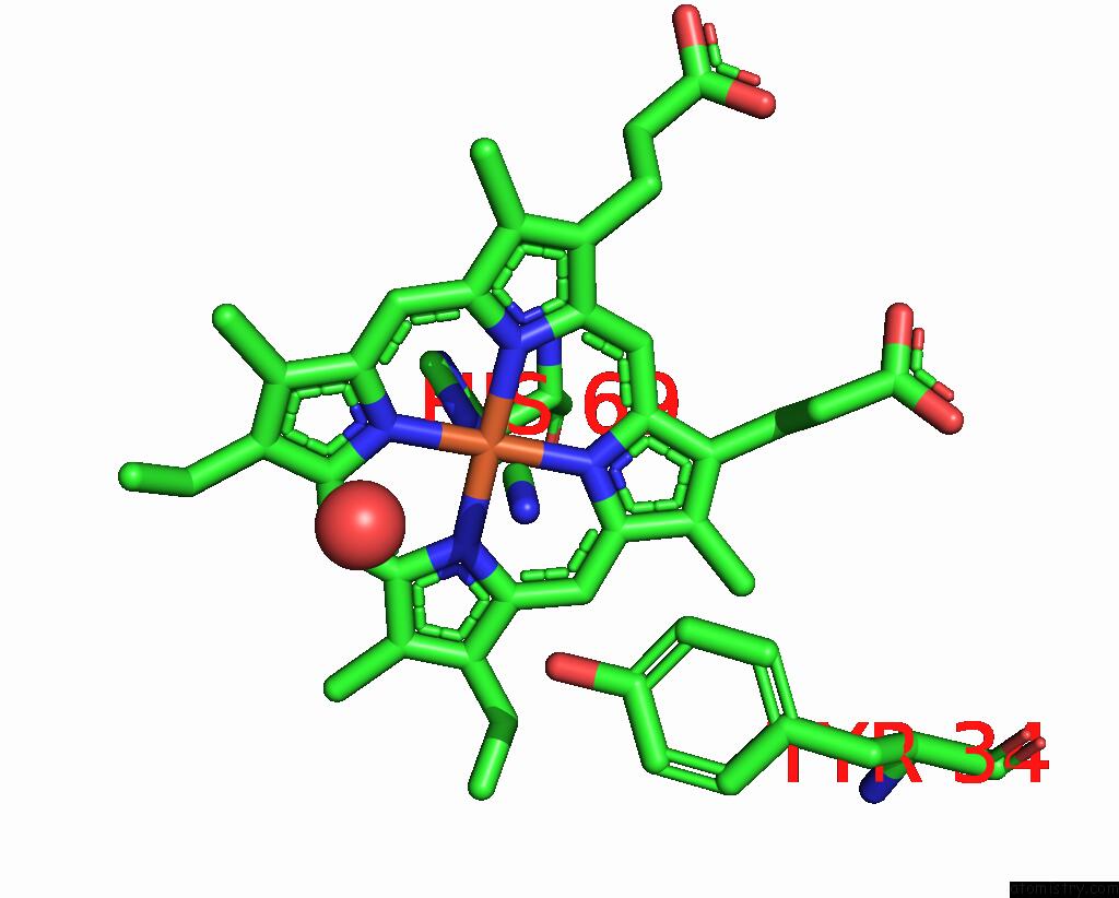
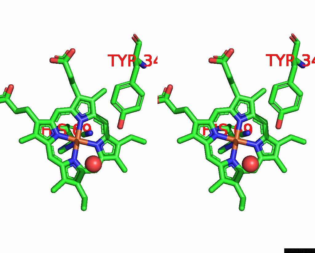
Iron binding site 1 out of 4 in 8ugz
Go back to
Iron binding site 1 out
of 4 in the Crystal Structure of Shewanella Benthica Group 1 Truncated Hemoglobin C51S C71S Variant
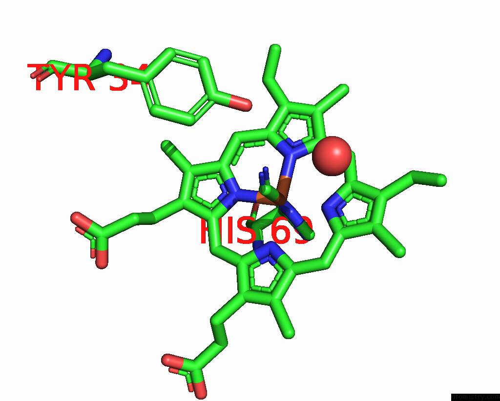
Mono view
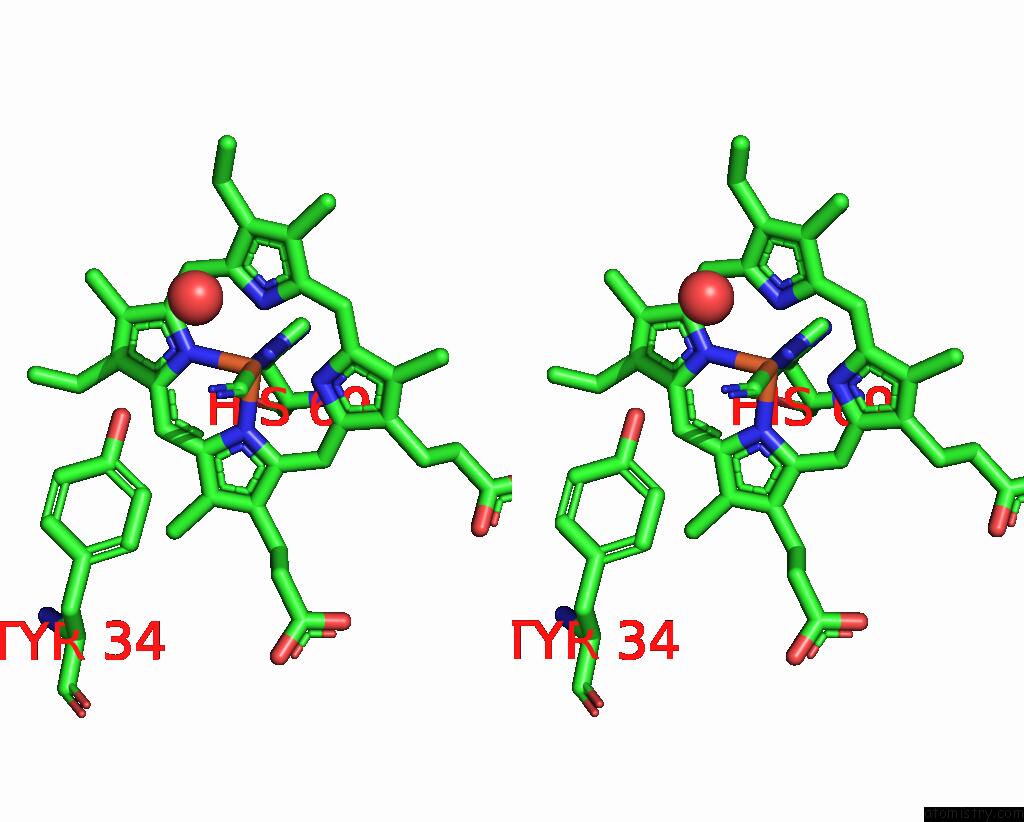
Stereo pair view
Mono view
Stereo pair view
A full contact list of Iron with other atoms in the Fe binding
site number 1 of Crystal Structure of Shewanella Benthica Group 1 Truncated Hemoglobin C51S C71S Variant within 5.0Å range:
|
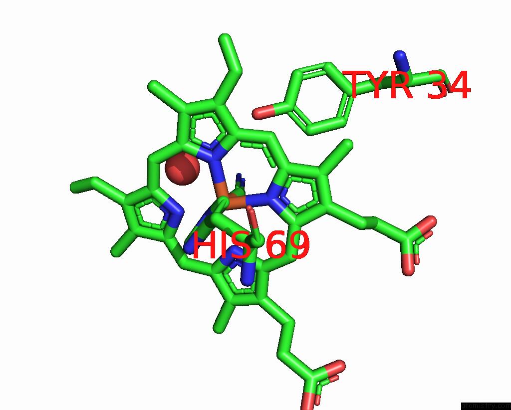
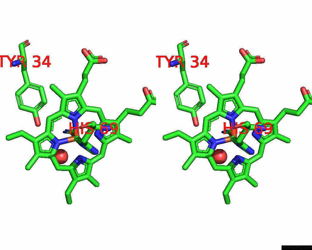
Iron binding site 2 out of 4 in 8ugz
Go back to
Iron binding site 2 out
of 4 in the Crystal Structure of Shewanella Benthica Group 1 Truncated Hemoglobin C51S C71S Variant
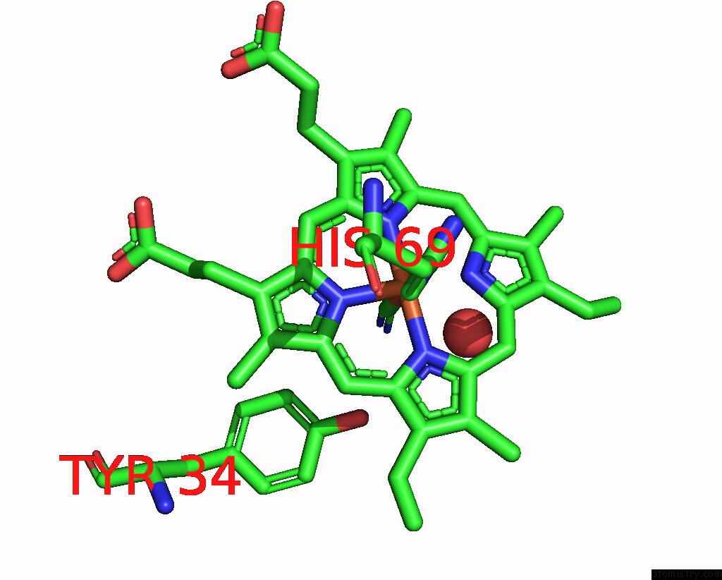
Mono view
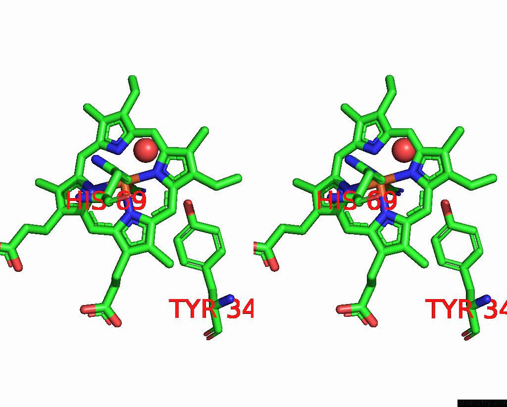
Stereo pair view
Mono view
Stereo pair view
A full contact list of Iron with other atoms in the Fe binding
site number 2 of Crystal Structure of Shewanella Benthica Group 1 Truncated Hemoglobin C51S C71S Variant within 5.0Å range:
|
Iron binding site 3 out of 4 in 8ugz
Go back to
Iron binding site 3 out
of 4 in the Crystal Structure of Shewanella Benthica Group 1 Truncated Hemoglobin C51S C71S Variant
Mono view
Stereo pair view
Mono view
Stereo pair view
A full contact list of Iron with other atoms in the Fe binding
site number 3 of Crystal Structure of Shewanella Benthica Group 1 Truncated Hemoglobin C51S C71S Variant within 5.0Å range:
|
Iron binding site 4 out of 4 in 8ugz
Go back to
Iron binding site 4 out
of 4 in the Crystal Structure of Shewanella Benthica Group 1 Truncated Hemoglobin C51S C71S Variant
Mono view
Stereo pair view
Mono view
Stereo pair view
A full contact list of Iron with other atoms in the Fe binding
site number 4 of Crystal Structure of Shewanella Benthica Group 1 Truncated Hemoglobin C51S C71S Variant within 5.0Å range:
|
Reference:
T.D.Schultz,
J.E.Martinez,
M.A.Siegler,
J.L.Schlessman,
J.T.J.Lecomte.
Crystal Structure of Shewanella Benthica Group 1 Truncated Hemoglobin C51S C71S Variant To Be Published.
Page generated: Sat Aug 10 18:36:50 2024
Last articles
Zn in 9J0NZn in 9J0O
Zn in 9J0P
Zn in 9FJX
Zn in 9EKB
Zn in 9C0F
Zn in 9CAH
Zn in 9CH0
Zn in 9CH3
Zn in 9CH1