Iron in PDB 8vpo: X-Ray Crystal Structure of Tige From Paramaledivibacter Caminithermalis
Protein crystallography data
The structure of X-Ray Crystal Structure of Tige From Paramaledivibacter Caminithermalis, PDB code: 8vpo
was solved by
T.L.Grove,
J.C.Lachowicz,
C.Zizola,
with X-Ray Crystallography technique. A brief refinement statistics is given in the table below:
Resolution Low / High (Å)
|
28.88 /
1.66
|
Space group
|
P 21 21 21
|
Cell size a, b, c (Å), α, β, γ (°)
|
69.172,
74.348,
91.765,
90,
90,
90
|
R / Rfree (%)
|
16.5 /
19.2
|
Iron Binding Sites:
The binding sites of Iron atom in the X-Ray Crystal Structure of Tige From Paramaledivibacter Caminithermalis
(pdb code 8vpo). This binding sites where shown within
5.0 Angstroms radius around Iron atom.
In total 8 binding sites of Iron where determined in the
X-Ray Crystal Structure of Tige From Paramaledivibacter Caminithermalis, PDB code: 8vpo:
Jump to Iron binding site number:
1;
2;
3;
4;
5;
6;
7;
8;
Iron binding site 1 out
of 8 in 8vpo
Go back to
Iron Binding Sites List in 8vpo
Iron binding site 1 out
of 8 in the X-Ray Crystal Structure of Tige From Paramaledivibacter Caminithermalis
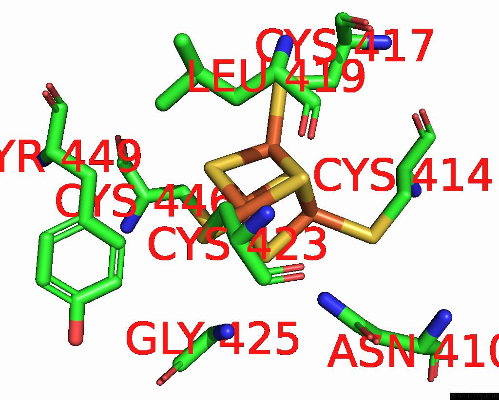
Mono view
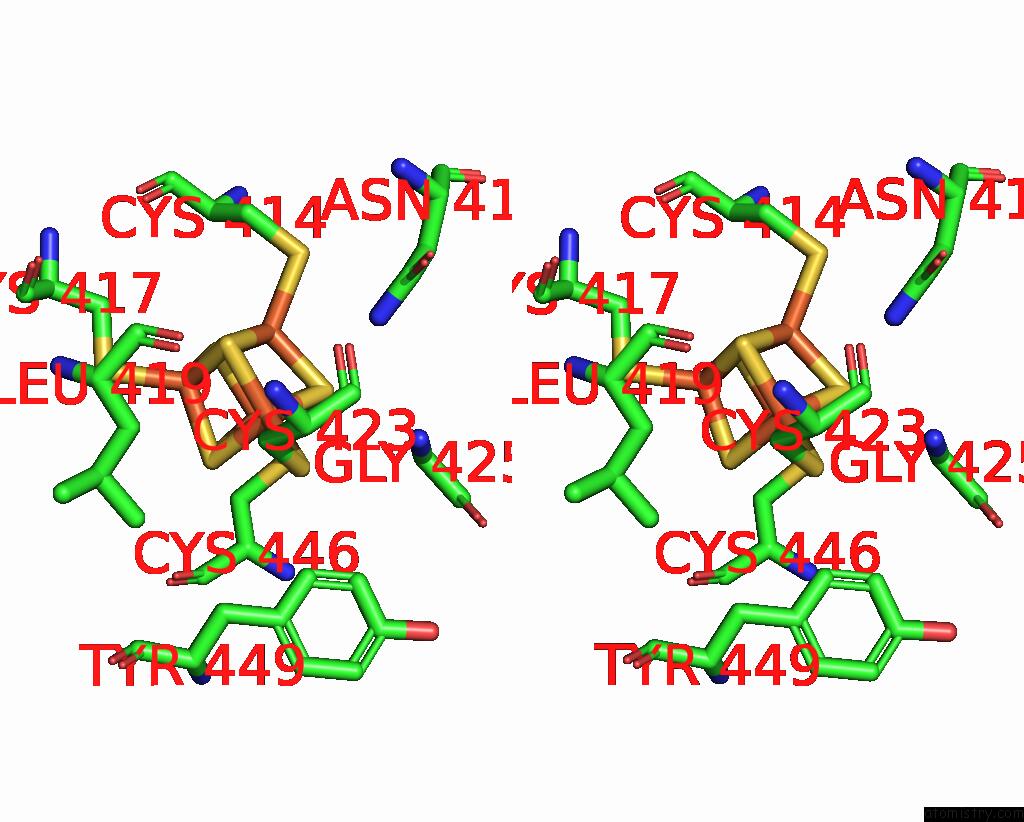
Stereo pair view
|
A full contact list of Iron with other atoms in the Fe binding
site number 1 of X-Ray Crystal Structure of Tige From Paramaledivibacter Caminithermalis within 5.0Å range:
probe
|
atom
|
residue
|
distance (Å)
|
B
|
Occ
|
A:Fe501
b:15.3
occ:1.00
|
FE1
|
A:SF4501
|
0.0
|
15.3
|
1.0
|
S4
|
A:SF4501
|
2.3
|
15.1
|
1.0
|
S3
|
A:SF4501
|
2.3
|
15.7
|
1.0
|
S2
|
A:SF4501
|
2.3
|
15.7
|
1.0
|
SG
|
A:CYS423
|
2.3
|
15.0
|
1.0
|
FE3
|
A:SF4501
|
2.7
|
16.1
|
1.0
|
FE4
|
A:SF4501
|
2.7
|
16.4
|
1.0
|
FE2
|
A:SF4501
|
2.8
|
15.9
|
1.0
|
CB
|
A:CYS423
|
3.2
|
15.6
|
1.0
|
S1
|
A:SF4501
|
3.9
|
16.9
|
1.0
|
ND2
|
A:ASN410
|
4.1
|
16.2
|
1.0
|
N
|
A:GLY425
|
4.2
|
15.0
|
1.0
|
O
|
A:CYS423
|
4.3
|
16.9
|
1.0
|
CA
|
A:GLY425
|
4.3
|
15.2
|
1.0
|
C
|
A:CYS423
|
4.3
|
15.8
|
1.0
|
CA
|
A:CYS423
|
4.4
|
15.2
|
1.0
|
CB
|
A:LEU419
|
4.5
|
17.9
|
1.0
|
CD2
|
A:TYR449
|
4.5
|
14.4
|
1.0
|
SG
|
A:CYS446
|
4.6
|
18.6
|
1.0
|
SG
|
A:CYS417
|
4.8
|
15.5
|
1.0
|
CD1
|
A:LEU419
|
4.8
|
16.8
|
1.0
|
SG
|
A:CYS414
|
4.9
|
17.9
|
1.0
|
O
|
A:LEU419
|
4.9
|
15.8
|
1.0
|
|
Iron binding site 2 out
of 8 in 8vpo
Go back to
Iron Binding Sites List in 8vpo
Iron binding site 2 out
of 8 in the X-Ray Crystal Structure of Tige From Paramaledivibacter Caminithermalis
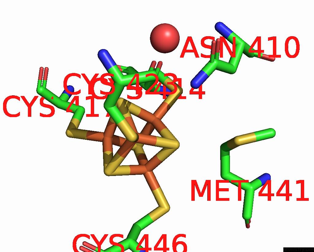
Mono view
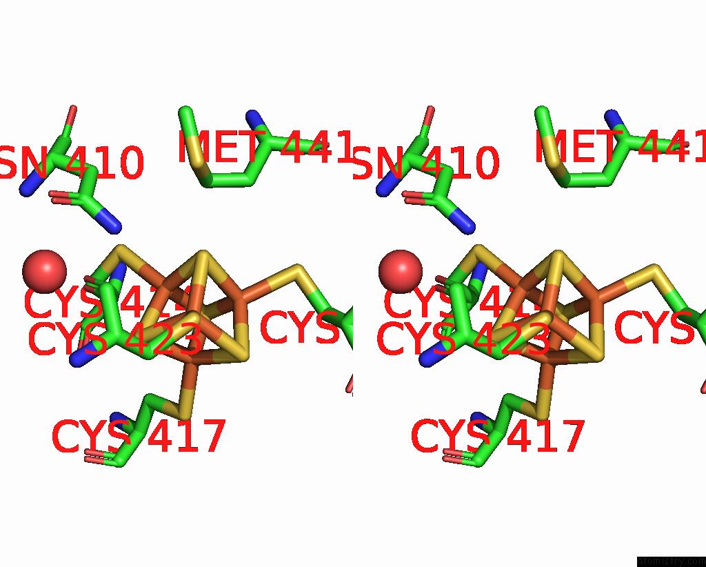
Stereo pair view
|
A full contact list of Iron with other atoms in the Fe binding
site number 2 of X-Ray Crystal Structure of Tige From Paramaledivibacter Caminithermalis within 5.0Å range:
probe
|
atom
|
residue
|
distance (Å)
|
B
|
Occ
|
A:Fe501
b:15.9
occ:1.00
|
FE2
|
A:SF4501
|
0.0
|
15.9
|
1.0
|
S3
|
A:SF4501
|
2.2
|
15.7
|
1.0
|
S1
|
A:SF4501
|
2.3
|
16.9
|
1.0
|
S4
|
A:SF4501
|
2.3
|
15.1
|
1.0
|
SG
|
A:CYS414
|
2.3
|
17.9
|
1.0
|
FE4
|
A:SF4501
|
2.7
|
16.4
|
1.0
|
FE3
|
A:SF4501
|
2.7
|
16.1
|
1.0
|
FE1
|
A:SF4501
|
2.8
|
15.3
|
1.0
|
CB
|
A:CYS414
|
3.3
|
16.2
|
1.0
|
ND2
|
A:ASN410
|
3.8
|
16.2
|
1.0
|
CA
|
A:CYS414
|
3.8
|
15.4
|
1.0
|
S2
|
A:SF4501
|
3.9
|
15.7
|
1.0
|
CG
|
A:MET441
|
4.3
|
15.9
|
1.0
|
O
|
A:HOH811
|
4.4
|
18.1
|
1.0
|
N
|
A:CYS414
|
4.6
|
16.5
|
1.0
|
SD
|
A:MET441
|
4.6
|
17.3
|
1.0
|
SG
|
A:CYS417
|
4.7
|
15.5
|
1.0
|
CB
|
A:CYS417
|
4.7
|
15.0
|
1.0
|
SG
|
A:CYS446
|
4.7
|
18.6
|
1.0
|
CG
|
A:ASN410
|
4.8
|
14.2
|
1.0
|
SG
|
A:CYS423
|
4.9
|
15.0
|
1.0
|
CB
|
A:ASN410
|
4.9
|
13.6
|
1.0
|
|
Iron binding site 3 out
of 8 in 8vpo
Go back to
Iron Binding Sites List in 8vpo
Iron binding site 3 out
of 8 in the X-Ray Crystal Structure of Tige From Paramaledivibacter Caminithermalis
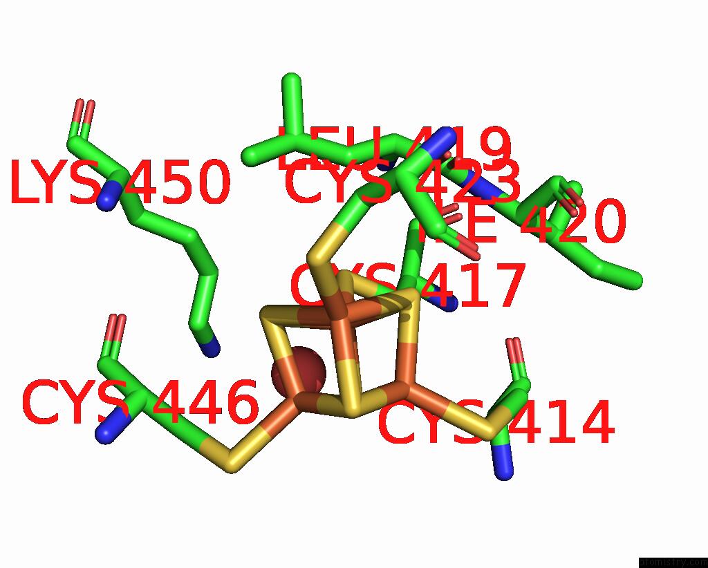
Mono view
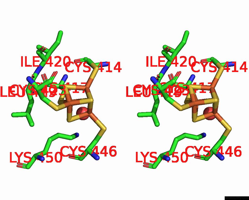
Stereo pair view
|
A full contact list of Iron with other atoms in the Fe binding
site number 3 of X-Ray Crystal Structure of Tige From Paramaledivibacter Caminithermalis within 5.0Å range:
probe
|
atom
|
residue
|
distance (Å)
|
B
|
Occ
|
A:Fe501
b:16.1
occ:1.00
|
FE3
|
A:SF4501
|
0.0
|
16.1
|
1.0
|
S2
|
A:SF4501
|
2.3
|
15.7
|
1.0
|
S4
|
A:SF4501
|
2.3
|
15.1
|
1.0
|
SG
|
A:CYS417
|
2.3
|
15.5
|
1.0
|
S1
|
A:SF4501
|
2.3
|
16.9
|
1.0
|
FE4
|
A:SF4501
|
2.7
|
16.4
|
1.0
|
FE2
|
A:SF4501
|
2.7
|
15.9
|
1.0
|
FE1
|
A:SF4501
|
2.7
|
15.3
|
1.0
|
CB
|
A:CYS417
|
3.2
|
15.0
|
1.0
|
S3
|
A:SF4501
|
3.8
|
15.7
|
1.0
|
O
|
A:HOH645
|
4.0
|
35.7
|
1.0
|
CB
|
A:LEU419
|
4.3
|
17.9
|
1.0
|
CA
|
A:CYS417
|
4.6
|
16.4
|
1.0
|
N
|
A:LEU419
|
4.6
|
15.7
|
1.0
|
SG
|
A:CYS414
|
4.8
|
17.9
|
1.0
|
CE
|
A:LYS450
|
4.8
|
32.4
|
1.0
|
SG
|
A:CYS423
|
4.8
|
15.0
|
1.0
|
CA
|
A:LEU419
|
4.8
|
14.3
|
1.0
|
C
|
A:LEU419
|
4.8
|
15.6
|
1.0
|
SG
|
A:CYS446
|
4.8
|
18.6
|
1.0
|
CG
|
A:LEU419
|
4.9
|
14.4
|
1.0
|
CD
|
A:LYS450
|
4.9
|
26.6
|
1.0
|
CB
|
A:CYS414
|
4.9
|
16.2
|
1.0
|
CD1
|
A:LEU419
|
4.9
|
16.8
|
1.0
|
N
|
A:ILE420
|
5.0
|
14.7
|
1.0
|
CA
|
A:CYS414
|
5.0
|
15.4
|
1.0
|
|
Iron binding site 4 out
of 8 in 8vpo
Go back to
Iron Binding Sites List in 8vpo
Iron binding site 4 out
of 8 in the X-Ray Crystal Structure of Tige From Paramaledivibacter Caminithermalis
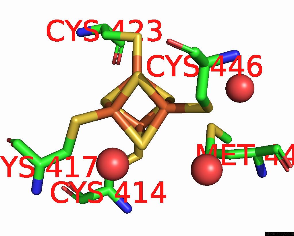
Mono view
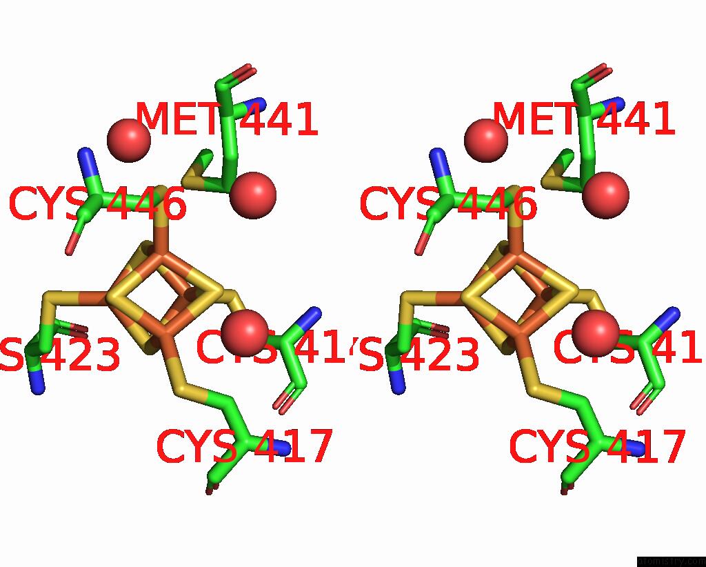
Stereo pair view
|
A full contact list of Iron with other atoms in the Fe binding
site number 4 of X-Ray Crystal Structure of Tige From Paramaledivibacter Caminithermalis within 5.0Å range:
probe
|
atom
|
residue
|
distance (Å)
|
B
|
Occ
|
A:Fe501
b:16.4
occ:1.00
|
FE4
|
A:SF4501
|
0.0
|
16.4
|
1.0
|
S1
|
A:SF4501
|
2.2
|
16.9
|
1.0
|
S3
|
A:SF4501
|
2.3
|
15.7
|
1.0
|
S2
|
A:SF4501
|
2.3
|
15.7
|
1.0
|
SG
|
A:CYS446
|
2.3
|
18.6
|
1.0
|
FE2
|
A:SF4501
|
2.7
|
15.9
|
1.0
|
FE3
|
A:SF4501
|
2.7
|
16.1
|
1.0
|
FE1
|
A:SF4501
|
2.7
|
15.3
|
1.0
|
CB
|
A:CYS446
|
3.1
|
16.5
|
1.0
|
S4
|
A:SF4501
|
3.8
|
15.1
|
1.0
|
CA
|
A:CYS446
|
3.9
|
16.6
|
1.0
|
O
|
A:HOH1029
|
4.1
|
36.7
|
1.0
|
O
|
A:HOH885
|
4.6
|
22.3
|
1.0
|
CG
|
A:MET441
|
4.6
|
15.9
|
1.0
|
CB
|
A:MET441
|
4.7
|
16.8
|
1.0
|
SG
|
A:CYS423
|
4.7
|
15.0
|
1.0
|
SG
|
A:CYS417
|
4.7
|
15.5
|
1.0
|
SG
|
A:CYS414
|
4.8
|
17.9
|
1.0
|
O
|
A:HOH645
|
4.9
|
35.7
|
1.0
|
N
|
A:CYS446
|
4.9
|
16.5
|
1.0
|
|
Iron binding site 5 out
of 8 in 8vpo
Go back to
Iron Binding Sites List in 8vpo
Iron binding site 5 out
of 8 in the X-Ray Crystal Structure of Tige From Paramaledivibacter Caminithermalis
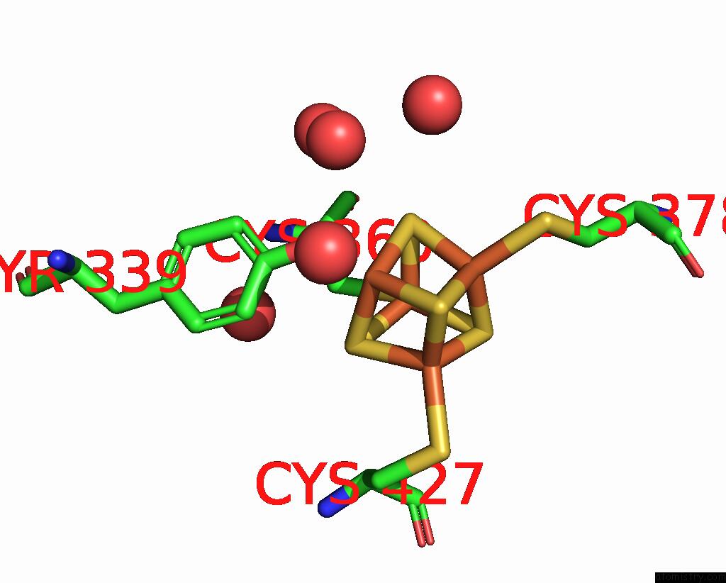
Mono view
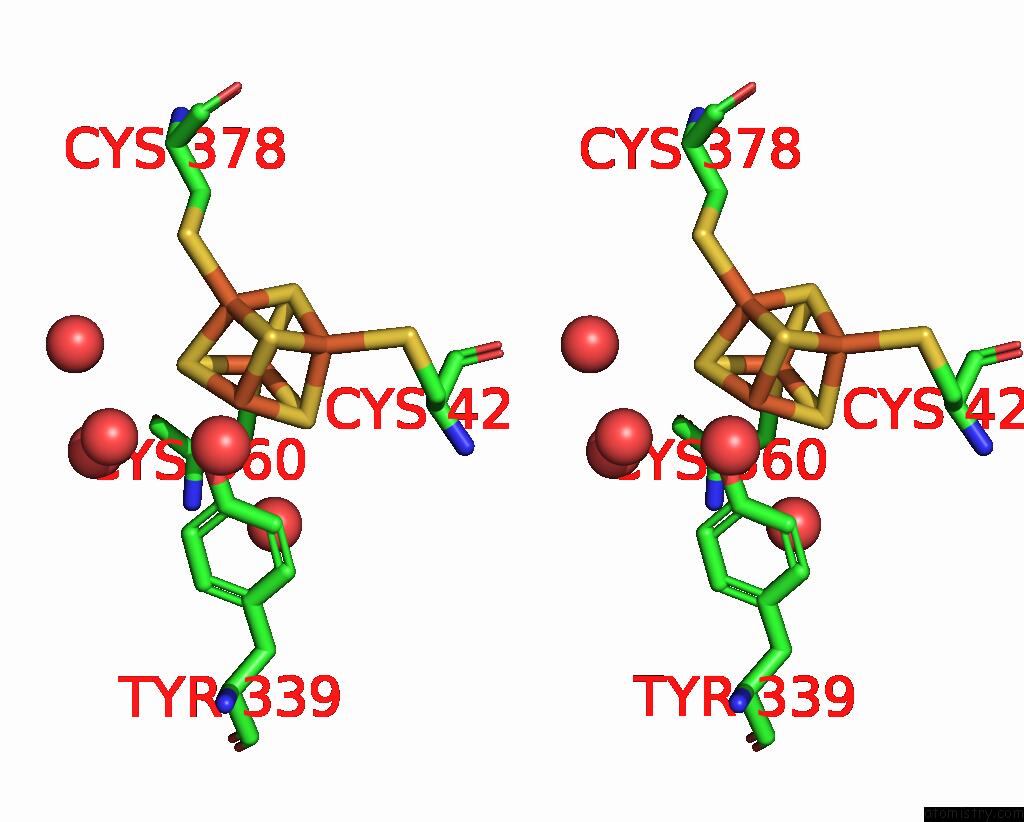
Stereo pair view
|
A full contact list of Iron with other atoms in the Fe binding
site number 5 of X-Ray Crystal Structure of Tige From Paramaledivibacter Caminithermalis within 5.0Å range:
probe
|
atom
|
residue
|
distance (Å)
|
B
|
Occ
|
A:Fe502
b:18.2
occ:1.00
|
FE1
|
A:SF4502
|
0.0
|
18.2
|
1.0
|
OH
|
A:TYR339
|
2.0
|
20.1
|
1.0
|
S2
|
A:SF4502
|
2.3
|
17.3
|
1.0
|
S3
|
A:SF4502
|
2.3
|
17.8
|
1.0
|
S4
|
A:SF4502
|
2.3
|
18.7
|
1.0
|
FE2
|
A:SF4502
|
2.6
|
18.3
|
1.0
|
FE3
|
A:SF4502
|
2.7
|
17.4
|
1.0
|
FE4
|
A:SF4502
|
2.7
|
17.4
|
1.0
|
CZ
|
A:TYR339
|
2.9
|
18.3
|
1.0
|
CE2
|
A:TYR339
|
3.6
|
20.3
|
1.0
|
O
|
A:HOH763
|
3.8
|
33.6
|
1.0
|
S1
|
A:SF4502
|
3.8
|
17.5
|
1.0
|
O
|
A:HOH633
|
3.8
|
32.0
|
1.0
|
CE1
|
A:TYR339
|
3.9
|
18.1
|
1.0
|
O
|
A:HOH660
|
4.6
|
20.8
|
1.0
|
SG
|
A:CYS378
|
4.7
|
17.6
|
1.0
|
O
|
A:HOH873
|
4.7
|
28.4
|
1.0
|
O
|
A:HOH874
|
4.7
|
36.9
|
1.0
|
CD2
|
A:TYR339
|
4.8
|
15.1
|
1.0
|
SG
|
A:CYS427
|
4.8
|
19.6
|
1.0
|
SG
|
A:CYS360
|
4.8
|
17.7
|
1.0
|
|
Iron binding site 6 out
of 8 in 8vpo
Go back to
Iron Binding Sites List in 8vpo
Iron binding site 6 out
of 8 in the X-Ray Crystal Structure of Tige From Paramaledivibacter Caminithermalis
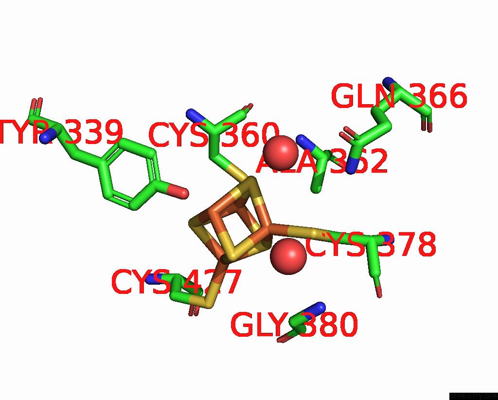
Mono view
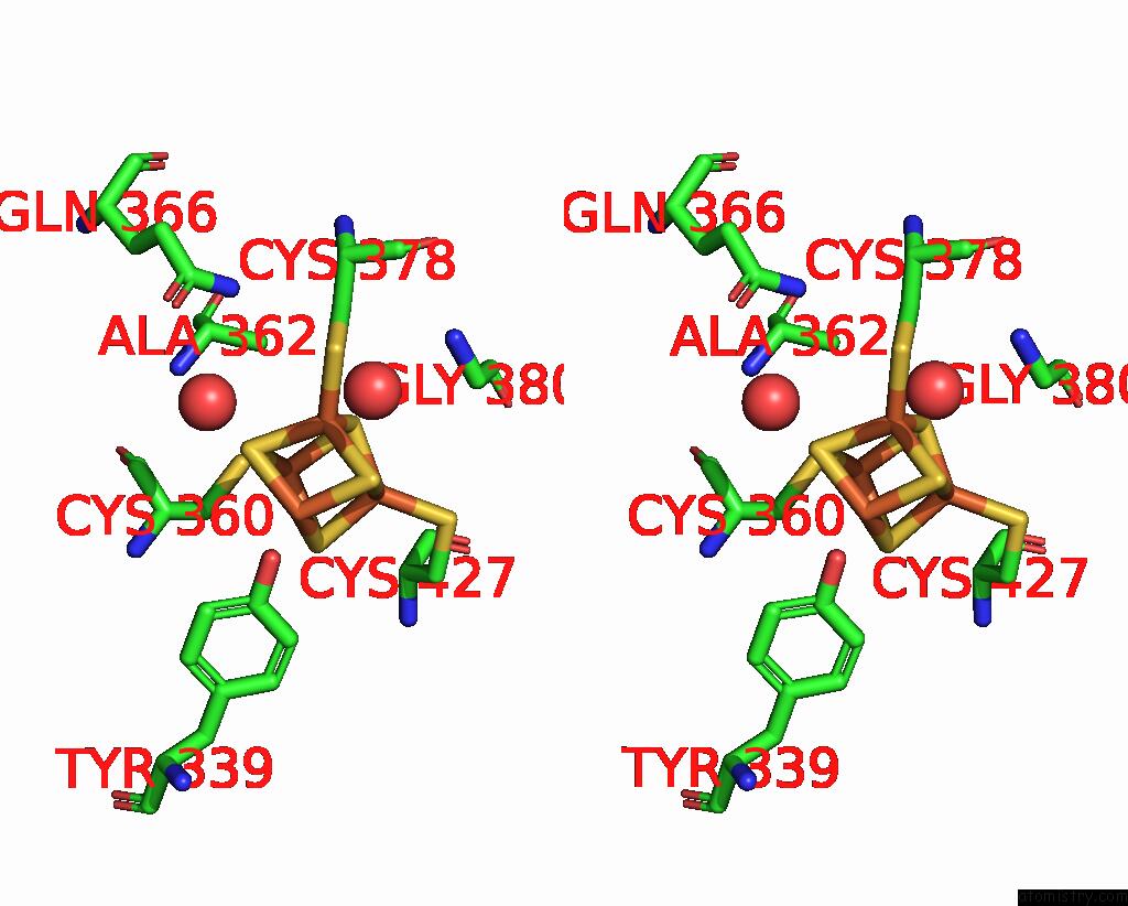
Stereo pair view
|
A full contact list of Iron with other atoms in the Fe binding
site number 6 of X-Ray Crystal Structure of Tige From Paramaledivibacter Caminithermalis within 5.0Å range:
probe
|
atom
|
residue
|
distance (Å)
|
B
|
Occ
|
A:Fe502
b:18.3
occ:1.00
|
FE2
|
A:SF4502
|
0.0
|
18.3
|
1.0
|
S1
|
A:SF4502
|
2.3
|
17.5
|
1.0
|
S3
|
A:SF4502
|
2.3
|
17.8
|
1.0
|
S4
|
A:SF4502
|
2.3
|
18.7
|
1.0
|
SG
|
A:CYS378
|
2.3
|
17.6
|
1.0
|
FE1
|
A:SF4502
|
2.6
|
18.2
|
1.0
|
FE3
|
A:SF4502
|
2.7
|
17.4
|
1.0
|
FE4
|
A:SF4502
|
2.7
|
17.4
|
1.0
|
CB
|
A:CYS378
|
3.3
|
17.3
|
1.0
|
S2
|
A:SF4502
|
3.8
|
17.3
|
1.0
|
O
|
A:HOH898
|
4.3
|
27.9
|
1.0
|
CB
|
A:ALA362
|
4.4
|
18.0
|
1.0
|
CA
|
A:GLY380
|
4.4
|
21.6
|
1.0
|
OH
|
A:TYR339
|
4.4
|
20.1
|
1.0
|
O
|
A:HOH874
|
4.5
|
36.9
|
1.0
|
N
|
A:GLY380
|
4.5
|
19.4
|
1.0
|
NE2
|
A:GLN366
|
4.7
|
19.4
|
1.0
|
CA
|
A:CYS378
|
4.7
|
18.1
|
1.0
|
SG
|
A:CYS360
|
4.8
|
17.7
|
1.0
|
SG
|
A:CYS427
|
4.9
|
19.6
|
1.0
|
C
|
A:GLY380
|
4.9
|
23.0
|
1.0
|
|
Iron binding site 7 out
of 8 in 8vpo
Go back to
Iron Binding Sites List in 8vpo
Iron binding site 7 out
of 8 in the X-Ray Crystal Structure of Tige From Paramaledivibacter Caminithermalis
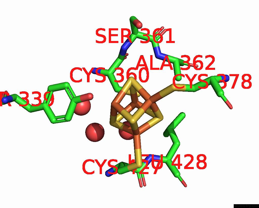
Mono view
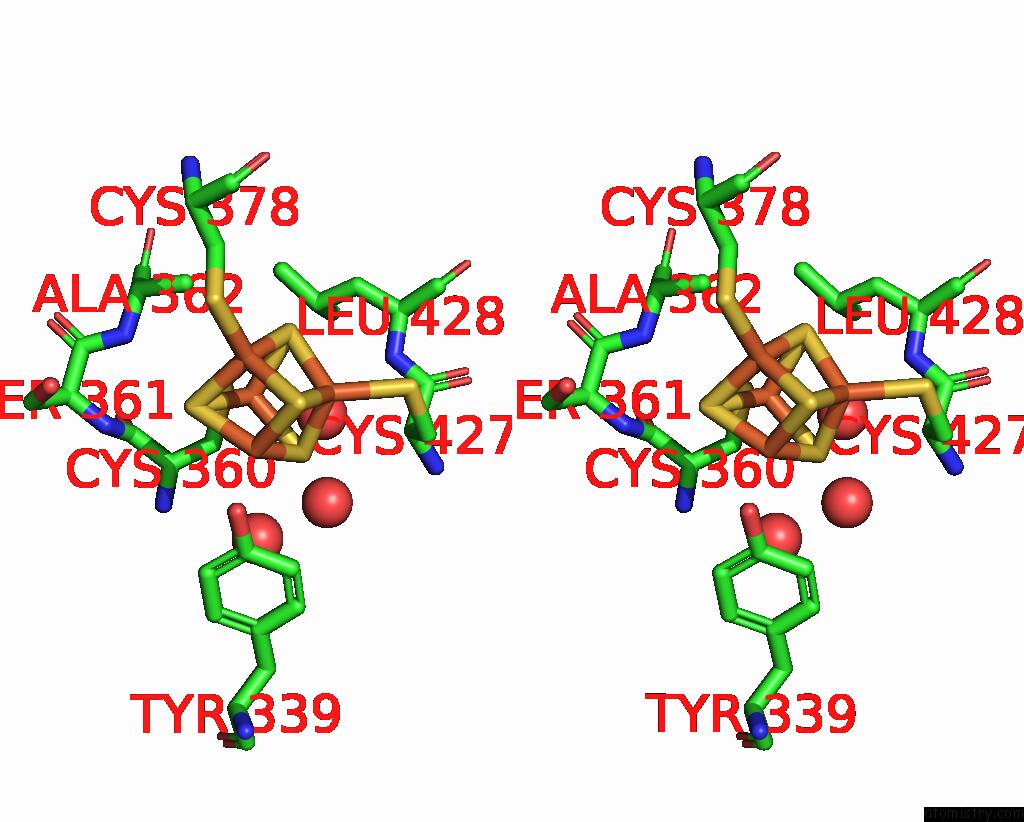
Stereo pair view
|
A full contact list of Iron with other atoms in the Fe binding
site number 7 of X-Ray Crystal Structure of Tige From Paramaledivibacter Caminithermalis within 5.0Å range:
probe
|
atom
|
residue
|
distance (Å)
|
B
|
Occ
|
A:Fe502
b:17.4
occ:1.00
|
FE3
|
A:SF4502
|
0.0
|
17.4
|
1.0
|
S1
|
A:SF4502
|
2.3
|
17.5
|
1.0
|
S4
|
A:SF4502
|
2.3
|
18.7
|
1.0
|
S2
|
A:SF4502
|
2.3
|
17.3
|
1.0
|
SG
|
A:CYS360
|
2.4
|
17.7
|
1.0
|
FE1
|
A:SF4502
|
2.7
|
18.2
|
1.0
|
FE2
|
A:SF4502
|
2.7
|
18.3
|
1.0
|
FE4
|
A:SF4502
|
2.8
|
17.4
|
1.0
|
CB
|
A:CYS360
|
3.4
|
15.4
|
1.0
|
O
|
A:HOH893
|
3.8
|
21.6
|
1.0
|
CA
|
A:CYS360
|
3.9
|
15.4
|
1.0
|
S3
|
A:SF4502
|
3.9
|
17.8
|
1.0
|
CB
|
A:ALA362
|
4.0
|
18.0
|
1.0
|
N
|
A:ALA362
|
4.4
|
16.2
|
1.0
|
C
|
A:CYS360
|
4.5
|
17.7
|
1.0
|
OH
|
A:TYR339
|
4.5
|
20.1
|
1.0
|
O
|
A:HOH660
|
4.5
|
20.8
|
1.0
|
CG
|
A:LEU428
|
4.6
|
19.2
|
1.0
|
N
|
A:SER361
|
4.6
|
19.2
|
1.0
|
O
|
A:HOH671
|
4.7
|
24.8
|
1.0
|
N
|
A:LEU428
|
4.8
|
15.4
|
1.0
|
SG
|
A:CYS378
|
4.8
|
17.6
|
1.0
|
SG
|
A:CYS427
|
4.8
|
19.6
|
1.0
|
CA
|
A:ALA362
|
4.9
|
13.6
|
1.0
|
CD1
|
A:LEU428
|
4.9
|
20.0
|
1.0
|
|
Iron binding site 8 out
of 8 in 8vpo
Go back to
Iron Binding Sites List in 8vpo
Iron binding site 8 out
of 8 in the X-Ray Crystal Structure of Tige From Paramaledivibacter Caminithermalis
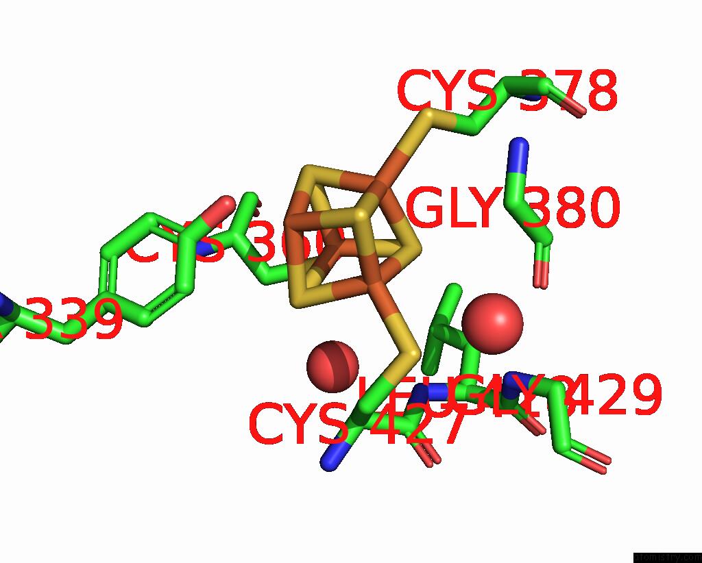
Mono view
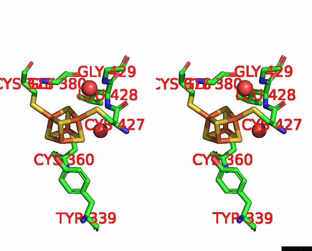
Stereo pair view
|
A full contact list of Iron with other atoms in the Fe binding
site number 8 of X-Ray Crystal Structure of Tige From Paramaledivibacter Caminithermalis within 5.0Å range:
probe
|
atom
|
residue
|
distance (Å)
|
B
|
Occ
|
A:Fe502
b:17.4
occ:1.00
|
FE4
|
A:SF4502
|
0.0
|
17.4
|
1.0
|
S3
|
A:SF4502
|
2.3
|
17.8
|
1.0
|
S2
|
A:SF4502
|
2.3
|
17.3
|
1.0
|
S1
|
A:SF4502
|
2.3
|
17.5
|
1.0
|
SG
|
A:CYS427
|
2.3
|
19.6
|
1.0
|
FE1
|
A:SF4502
|
2.7
|
18.2
|
1.0
|
FE2
|
A:SF4502
|
2.7
|
18.3
|
1.0
|
FE3
|
A:SF4502
|
2.8
|
17.4
|
1.0
|
CB
|
A:CYS427
|
3.4
|
20.1
|
1.0
|
CA
|
A:CYS427
|
3.9
|
15.7
|
1.0
|
S4
|
A:SF4502
|
3.9
|
18.7
|
1.0
|
N
|
A:LEU428
|
4.1
|
15.4
|
1.0
|
C
|
A:CYS427
|
4.4
|
17.0
|
1.0
|
OH
|
A:TYR339
|
4.5
|
20.1
|
1.0
|
O
|
A:HOH635
|
4.5
|
28.6
|
1.0
|
O
|
A:GLY380
|
4.6
|
24.9
|
1.0
|
CA
|
A:GLY380
|
4.7
|
21.6
|
1.0
|
O
|
A:HOH893
|
4.7
|
21.6
|
1.0
|
SG
|
A:CYS378
|
4.8
|
17.6
|
1.0
|
C
|
A:GLY380
|
4.8
|
23.0
|
1.0
|
N
|
A:GLY429
|
4.8
|
17.6
|
1.0
|
SG
|
A:CYS360
|
4.9
|
17.7
|
1.0
|
|
Reference:
Y.Lien,
J.C.Lachowicz,
A.Mendauletova,
C.Zizola,
T.Ngendahimana,
A.Kostenko,
S.S.Eaton,
J.A.Latham,
T.L.Grove.
Structural, Biochemical, and Bioinformatic Basis For Identifying Radical Sam Cyclopropyl Synthases Acs Chem.Biol. 2024.
ISSN: ESSN 1554-8937
DOI: 10.1021/ACSCHEMBIO.3C00583
Page generated: Sat Aug 10 18:45:43 2024
|