Iron in PDB 8vz7: Crystal Structure of Human Cytochrome P450 2C9*27 (R150L) Genetic Variant in Complex with the Drug Losartan
Enzymatic activity of Crystal Structure of Human Cytochrome P450 2C9*27 (R150L) Genetic Variant in Complex with the Drug Losartan
All present enzymatic activity of Crystal Structure of Human Cytochrome P450 2C9*27 (R150L) Genetic Variant in Complex with the Drug Losartan:
1.14.14.1; 1.14.14.51; 1.14.14.52; 1.14.14.53;
1.14.14.1; 1.14.14.51; 1.14.14.52; 1.14.14.53;
Protein crystallography data
The structure of Crystal Structure of Human Cytochrome P450 2C9*27 (R150L) Genetic Variant in Complex with the Drug Losartan, PDB code: 8vz7
was solved by
M.B.Shah,
with X-Ray Crystallography technique. A brief refinement statistics is given in the table below:
| Resolution Low / High (Å) | 50.01 / 2.53 |
| Space group | I 2 2 2 |
| Cell size a, b, c (Å), α, β, γ (°) | 74.89, 142.17, 163.07, 90, 90, 90 |
| R / Rfree (%) | 15.1 / 21.5 |
Other elements in 8vz7:
The structure of Crystal Structure of Human Cytochrome P450 2C9*27 (R150L) Genetic Variant in Complex with the Drug Losartan also contains other interesting chemical elements:
| Potassium | (K) | 1 atom |
| Chlorine | (Cl) | 2 atoms |
Iron Binding Sites:
The binding sites of Iron atom in the Crystal Structure of Human Cytochrome P450 2C9*27 (R150L) Genetic Variant in Complex with the Drug Losartan
(pdb code 8vz7). This binding sites where shown within
5.0 Angstroms radius around Iron atom.
In total only one binding site of Iron was determined in the Crystal Structure of Human Cytochrome P450 2C9*27 (R150L) Genetic Variant in Complex with the Drug Losartan, PDB code: 8vz7:
In total only one binding site of Iron was determined in the Crystal Structure of Human Cytochrome P450 2C9*27 (R150L) Genetic Variant in Complex with the Drug Losartan, PDB code: 8vz7:
Iron binding site 1 out of 1 in 8vz7
Go back to
Iron binding site 1 out
of 1 in the Crystal Structure of Human Cytochrome P450 2C9*27 (R150L) Genetic Variant in Complex with the Drug Losartan
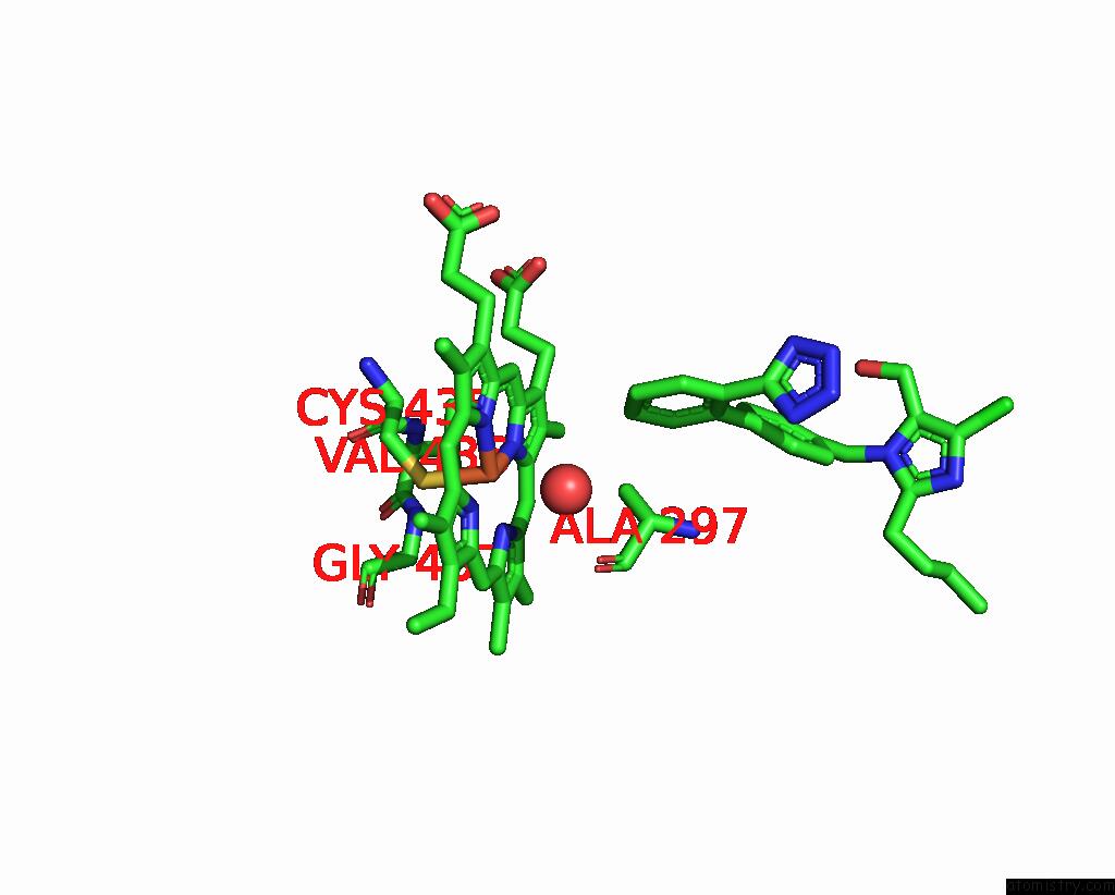
Mono view
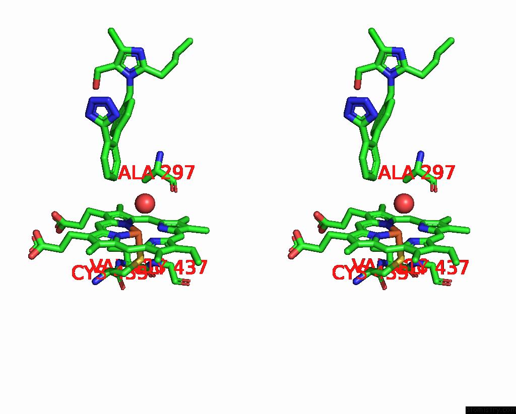
Stereo pair view
Mono view
Stereo pair view
A full contact list of Iron with other atoms in the Fe binding
site number 1 of Crystal Structure of Human Cytochrome P450 2C9*27 (R150L) Genetic Variant in Complex with the Drug Losartan within 5.0Å range:
|
Reference:
S.J.Parikh,
S.Edara,
S.Deodhar,
A.K.Singh,
K.Maekawa,
Q.Zhang,
K.C.Glass,
M.B.Shah.
Structural and Biophysical Analysis of Cytochrome P450 2C9*14 and *27 Variants in Complex with Losartan J.Inorg.Biochem. 2024.
ISSN: ISSN 0162-0134
DOI: 10.1016/J.JINORGBIO.2024.112622
Page generated: Sat Aug 10 18:47:26 2024
ISSN: ISSN 0162-0134
DOI: 10.1016/J.JINORGBIO.2024.112622
Last articles
Zn in 9J0NZn in 9J0O
Zn in 9J0P
Zn in 9FJX
Zn in 9EKB
Zn in 9C0F
Zn in 9CAH
Zn in 9CH0
Zn in 9CH3
Zn in 9CH1