Iron in PDB 8w6y: Ferritin From Ureaplasma Diversum Soaking in FE2+ Solution For 10 Min
Protein crystallography data
The structure of Ferritin From Ureaplasma Diversum Soaking in FE2+ Solution For 10 Min, PDB code: 8w6y
was solved by
W.M.Wang,
H.F.Xi,
W.J.Gong,
D.Y.Ma,
H.F.Wang,
with X-Ray Crystallography technique. A brief refinement statistics is given in the table below:
| Resolution Low / High (Å) | 31.91 / 2.50 |
| Space group | F 4 3 2 |
| Cell size a, b, c (Å), α, β, γ (°) | 180.513, 180.513, 180.513, 90, 90, 90 |
| R / Rfree (%) | 19.7 / 24.7 |
Iron Binding Sites:
The binding sites of Iron atom in the Ferritin From Ureaplasma Diversum Soaking in FE2+ Solution For 10 Min
(pdb code 8w6y). This binding sites where shown within
5.0 Angstroms radius around Iron atom.
In total 8 binding sites of Iron where determined in the Ferritin From Ureaplasma Diversum Soaking in FE2+ Solution For 10 Min, PDB code: 8w6y:
Jump to Iron binding site number: 1; 2; 3; 4; 5; 6; 7; 8;
In total 8 binding sites of Iron where determined in the Ferritin From Ureaplasma Diversum Soaking in FE2+ Solution For 10 Min, PDB code: 8w6y:
Jump to Iron binding site number: 1; 2; 3; 4; 5; 6; 7; 8;
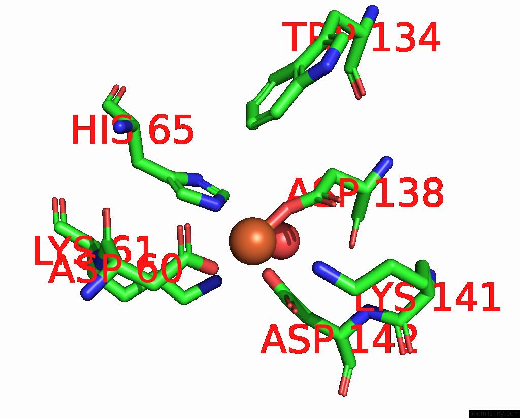
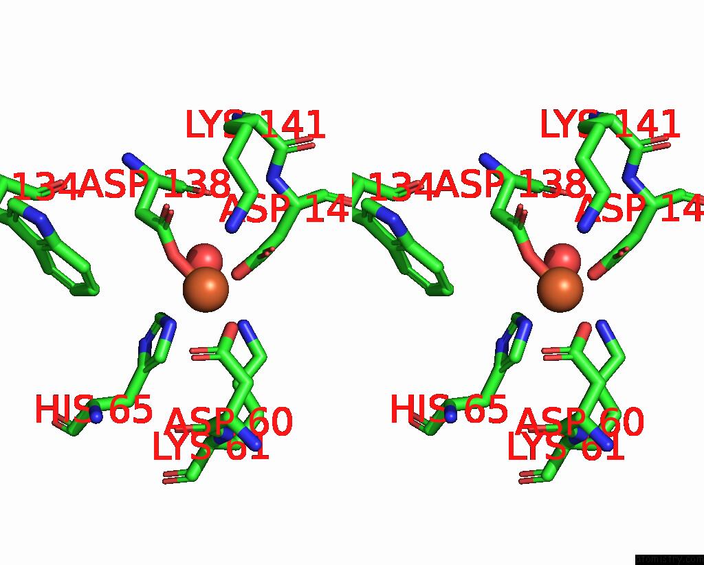
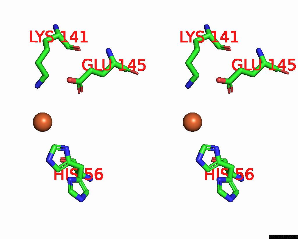
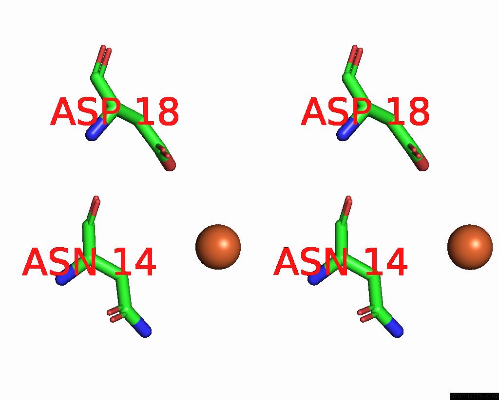
Iron binding site 1 out of 8 in 8w6y
Go back to
Iron binding site 1 out
of 8 in the Ferritin From Ureaplasma Diversum Soaking in FE2+ Solution For 10 Min
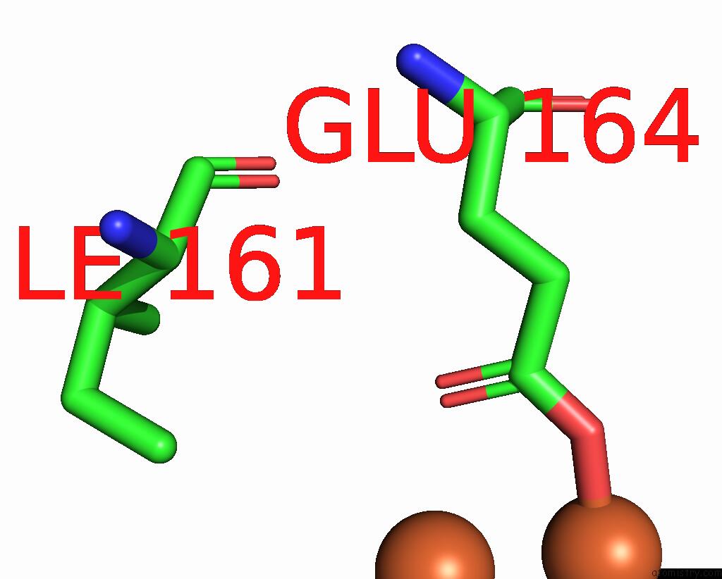
Mono view
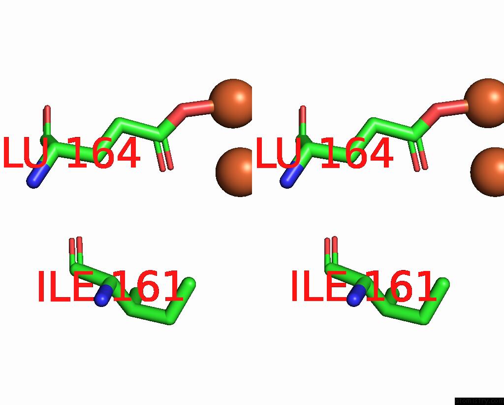
Stereo pair view
Mono view
Stereo pair view
A full contact list of Iron with other atoms in the Fe binding
site number 1 of Ferritin From Ureaplasma Diversum Soaking in FE2+ Solution For 10 Min within 5.0Å range:
|
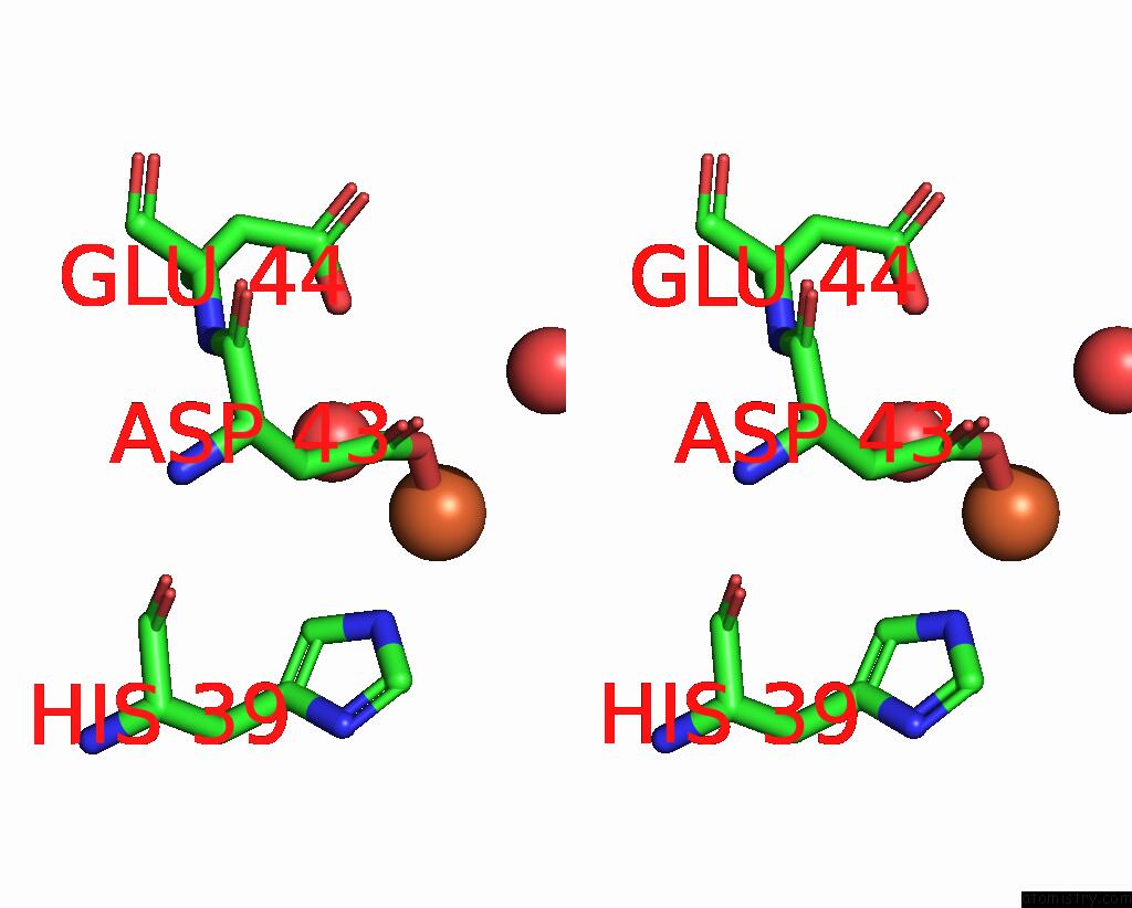
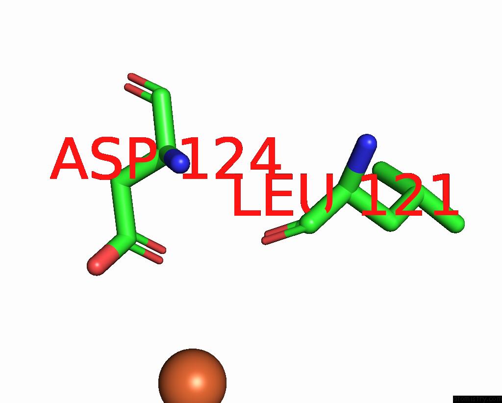
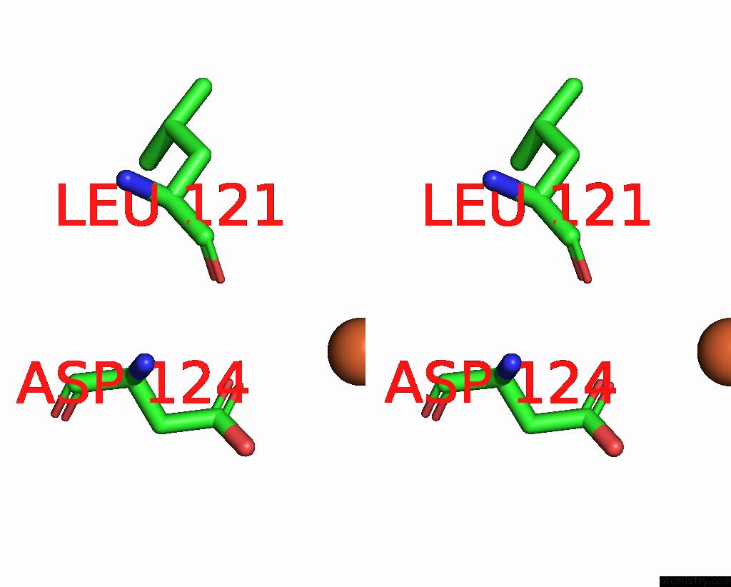
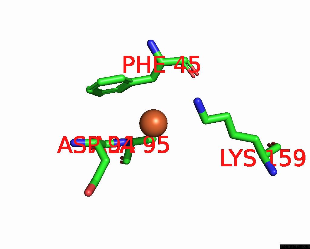
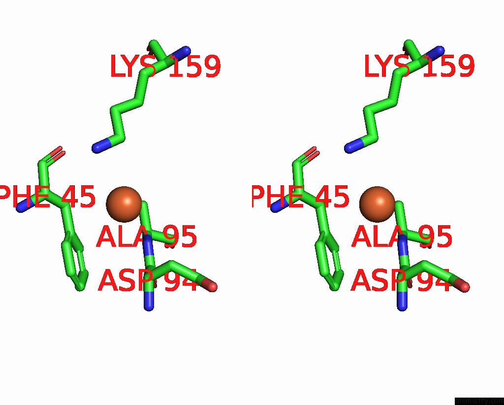
Iron binding site 2 out of 8 in 8w6y
Go back to
Iron binding site 2 out
of 8 in the Ferritin From Ureaplasma Diversum Soaking in FE2+ Solution For 10 Min
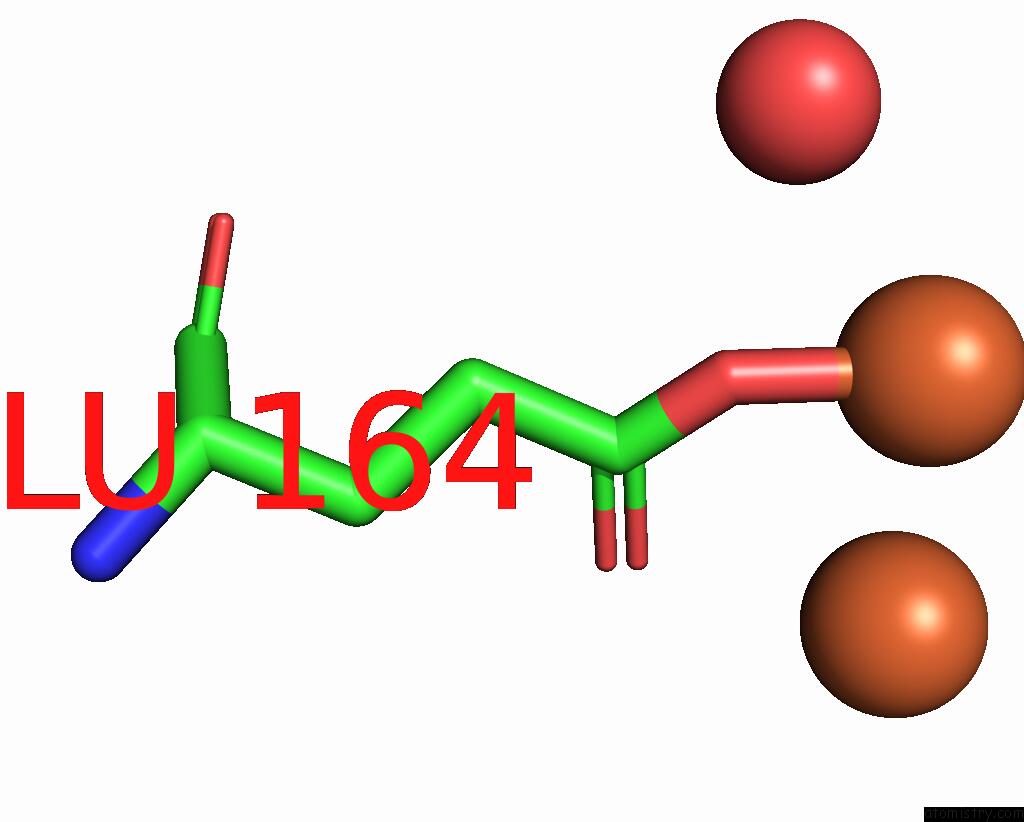
Mono view
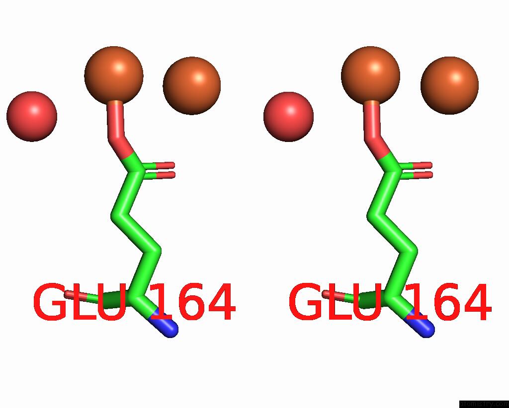
Stereo pair view
Mono view
Stereo pair view
A full contact list of Iron with other atoms in the Fe binding
site number 2 of Ferritin From Ureaplasma Diversum Soaking in FE2+ Solution For 10 Min within 5.0Å range:
|
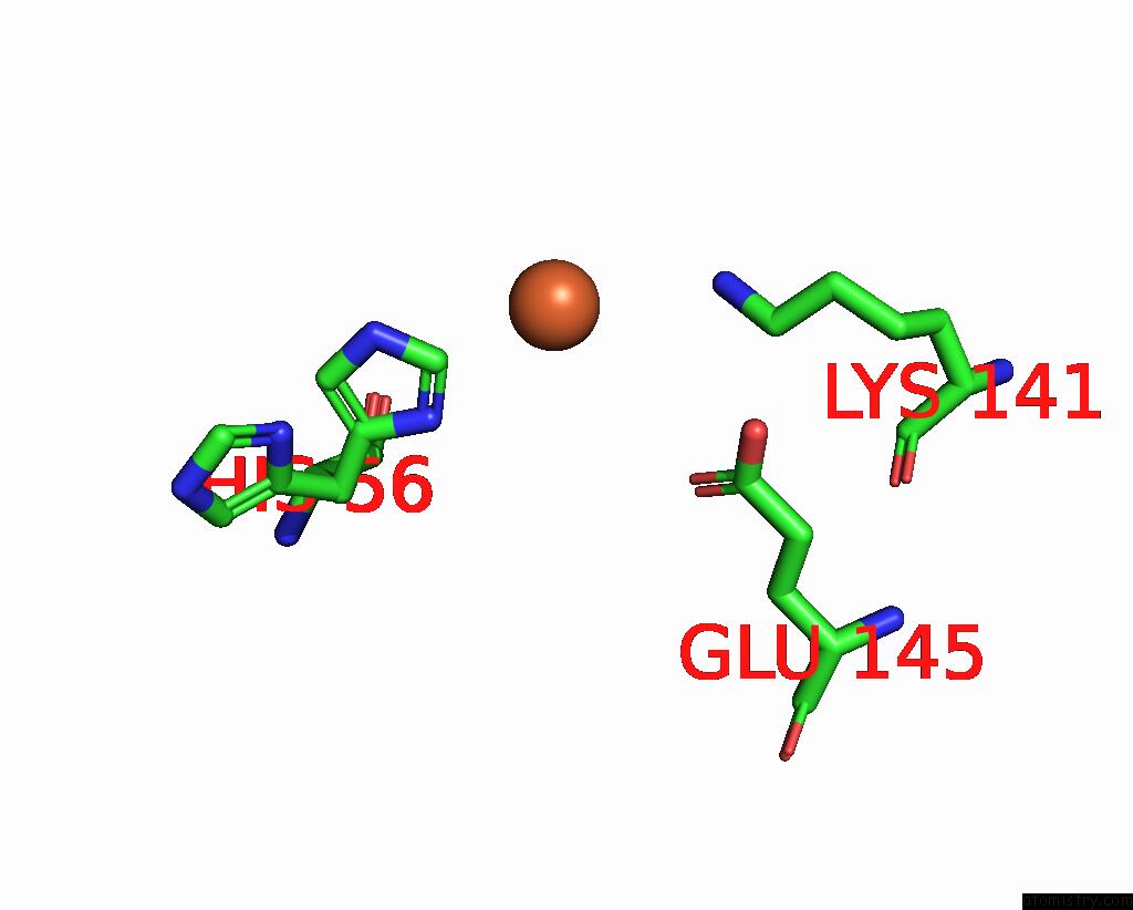
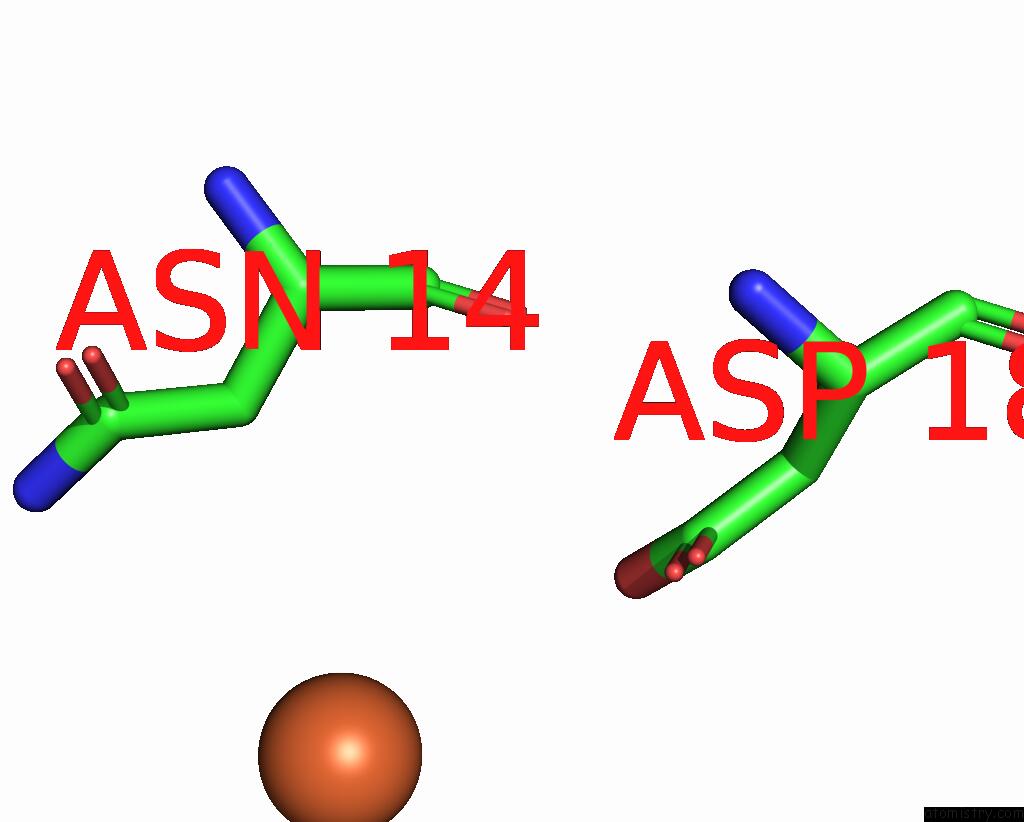
Iron binding site 3 out of 8 in 8w6y
Go back to
Iron binding site 3 out
of 8 in the Ferritin From Ureaplasma Diversum Soaking in FE2+ Solution For 10 Min
Mono view
Stereo pair view
Mono view
Stereo pair view
A full contact list of Iron with other atoms in the Fe binding
site number 3 of Ferritin From Ureaplasma Diversum Soaking in FE2+ Solution For 10 Min within 5.0Å range:
|
Iron binding site 4 out of 8 in 8w6y
Go back to
Iron binding site 4 out
of 8 in the Ferritin From Ureaplasma Diversum Soaking in FE2+ Solution For 10 Min

Mono view
Stereo pair view
Mono view
Stereo pair view
A full contact list of Iron with other atoms in the Fe binding
site number 4 of Ferritin From Ureaplasma Diversum Soaking in FE2+ Solution For 10 Min within 5.0Å range:
|
Iron binding site 5 out of 8 in 8w6y
Go back to
Iron binding site 5 out
of 8 in the Ferritin From Ureaplasma Diversum Soaking in FE2+ Solution For 10 Min
Mono view
Stereo pair view
Mono view
Stereo pair view
A full contact list of Iron with other atoms in the Fe binding
site number 5 of Ferritin From Ureaplasma Diversum Soaking in FE2+ Solution For 10 Min within 5.0Å range:
|
Iron binding site 6 out of 8 in 8w6y
Go back to
Iron binding site 6 out
of 8 in the Ferritin From Ureaplasma Diversum Soaking in FE2+ Solution For 10 Min
Mono view
Stereo pair view
Mono view
Stereo pair view
A full contact list of Iron with other atoms in the Fe binding
site number 6 of Ferritin From Ureaplasma Diversum Soaking in FE2+ Solution For 10 Min within 5.0Å range:
|
Iron binding site 7 out of 8 in 8w6y
Go back to
Iron binding site 7 out
of 8 in the Ferritin From Ureaplasma Diversum Soaking in FE2+ Solution For 10 Min
Mono view
Stereo pair view
Mono view
Stereo pair view
A full contact list of Iron with other atoms in the Fe binding
site number 7 of Ferritin From Ureaplasma Diversum Soaking in FE2+ Solution For 10 Min within 5.0Å range:
|
Iron binding site 8 out of 8 in 8w6y
Go back to
Iron binding site 8 out
of 8 in the Ferritin From Ureaplasma Diversum Soaking in FE2+ Solution For 10 Min
Mono view
Stereo pair view
Mono view
Stereo pair view
A full contact list of Iron with other atoms in the Fe binding
site number 8 of Ferritin From Ureaplasma Diversum Soaking in FE2+ Solution For 10 Min within 5.0Å range:
|
Reference:
W.Wang,
H.Xi,
D.Fu,
D.Ma,
W.Gong,
Y.Zhao,
X.Li,
L.Wu,
Y.Guo,
G.Zhao,
H.Wang.
Growth Process of Fe-O Nanoclusters with Different Sizes Biosynthesized By Protein Nanocages. J.Am.Chem.Soc. V. 146 11657 2024.
ISSN: ESSN 1520-5126
PubMed: 38641862
DOI: 10.1021/JACS.3C13830
Page generated: Sat Aug 10 18:54:37 2024
ISSN: ESSN 1520-5126
PubMed: 38641862
DOI: 10.1021/JACS.3C13830
Last articles
Zn in 9J0NZn in 9J0O
Zn in 9J0P
Zn in 9FJX
Zn in 9EKB
Zn in 9C0F
Zn in 9CAH
Zn in 9CH0
Zn in 9CH3
Zn in 9CH1