Iron »
PDB 8wgh-8xcg »
8wub »
Iron in PDB 8wub: The X-Ray Structure of Human Neuroglobin C120S Mutant
Protein crystallography data
The structure of The X-Ray Structure of Human Neuroglobin C120S Mutant, PDB code: 8wub
was solved by
Y.W.Lin,
H.Yuan,
with X-Ray Crystallography technique. A brief refinement statistics is given in the table below:
| Resolution Low / High (Å) | 28.52 / 1.50 |
| Space group | P 21 21 2 |
| Cell size a, b, c (Å), α, β, γ (°) | 84.009, 119.94, 38.857, 90, 90, 90 |
| R / Rfree (%) | 15.4 / 20.4 |
Iron Binding Sites:
The binding sites of Iron atom in the The X-Ray Structure of Human Neuroglobin C120S Mutant
(pdb code 8wub). This binding sites where shown within
5.0 Angstroms radius around Iron atom.
In total 3 binding sites of Iron where determined in the The X-Ray Structure of Human Neuroglobin C120S Mutant, PDB code: 8wub:
Jump to Iron binding site number: 1; 2; 3;
In total 3 binding sites of Iron where determined in the The X-Ray Structure of Human Neuroglobin C120S Mutant, PDB code: 8wub:
Jump to Iron binding site number: 1; 2; 3;
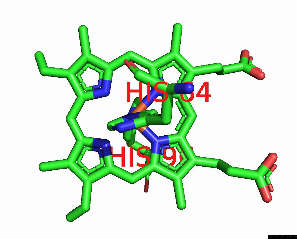
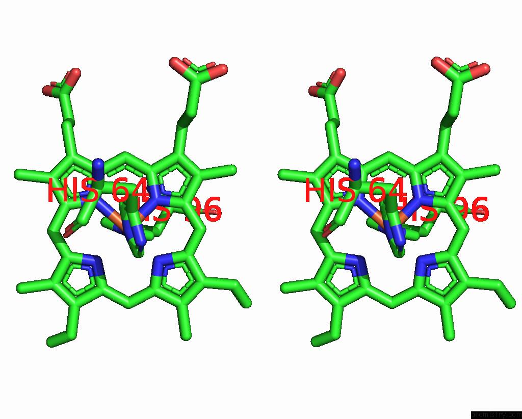
Iron binding site 1 out of 3 in 8wub
Go back to
Iron binding site 1 out
of 3 in the The X-Ray Structure of Human Neuroglobin C120S Mutant
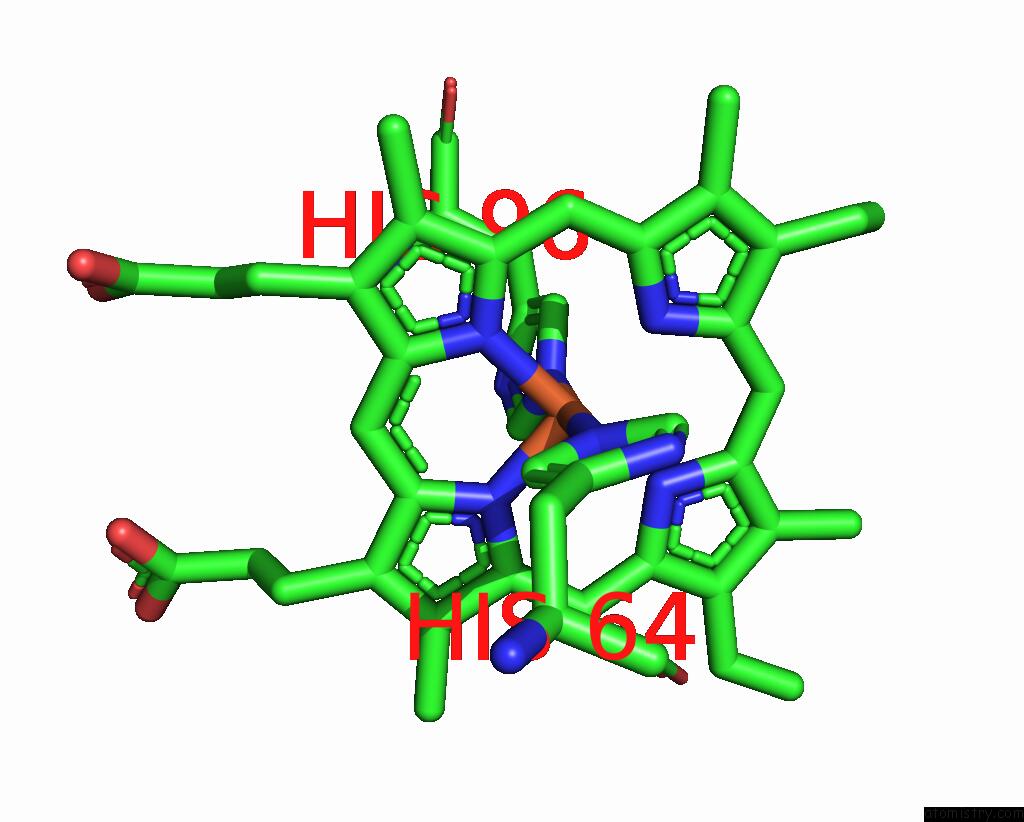
Mono view
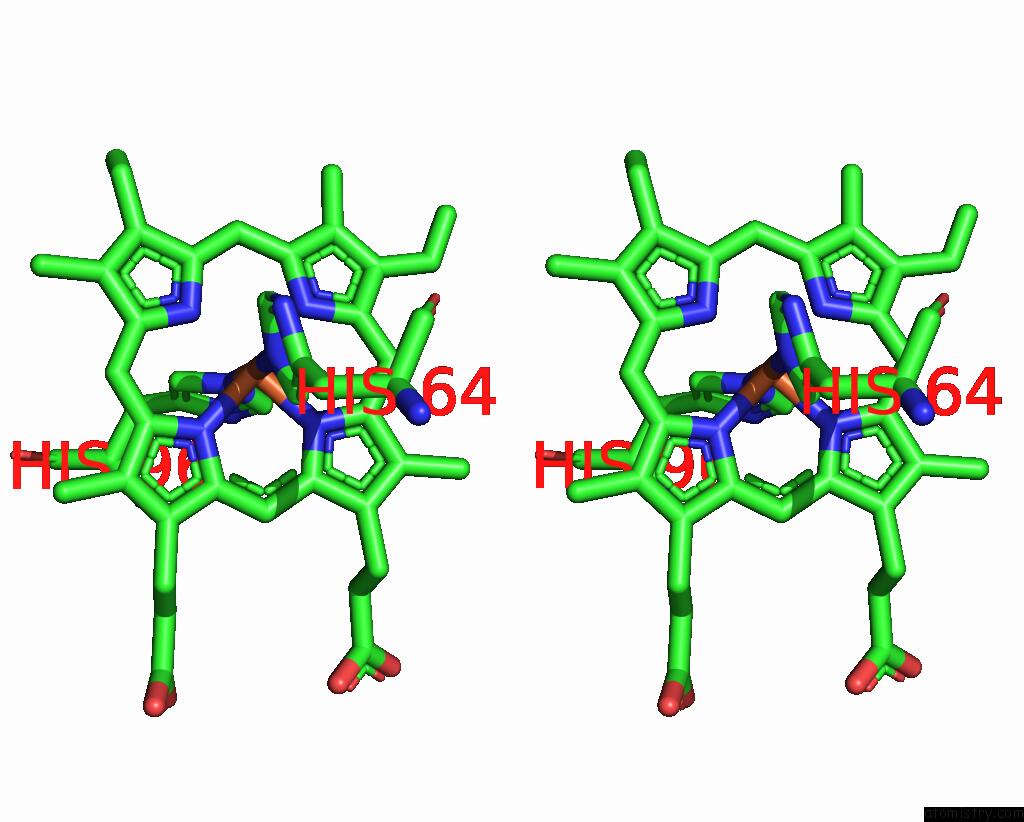
Stereo pair view
Mono view
Stereo pair view
A full contact list of Iron with other atoms in the Fe binding
site number 1 of The X-Ray Structure of Human Neuroglobin C120S Mutant within 5.0Å range:
|
Iron binding site 2 out of 3 in 8wub
Go back to
Iron binding site 2 out
of 3 in the The X-Ray Structure of Human Neuroglobin C120S Mutant
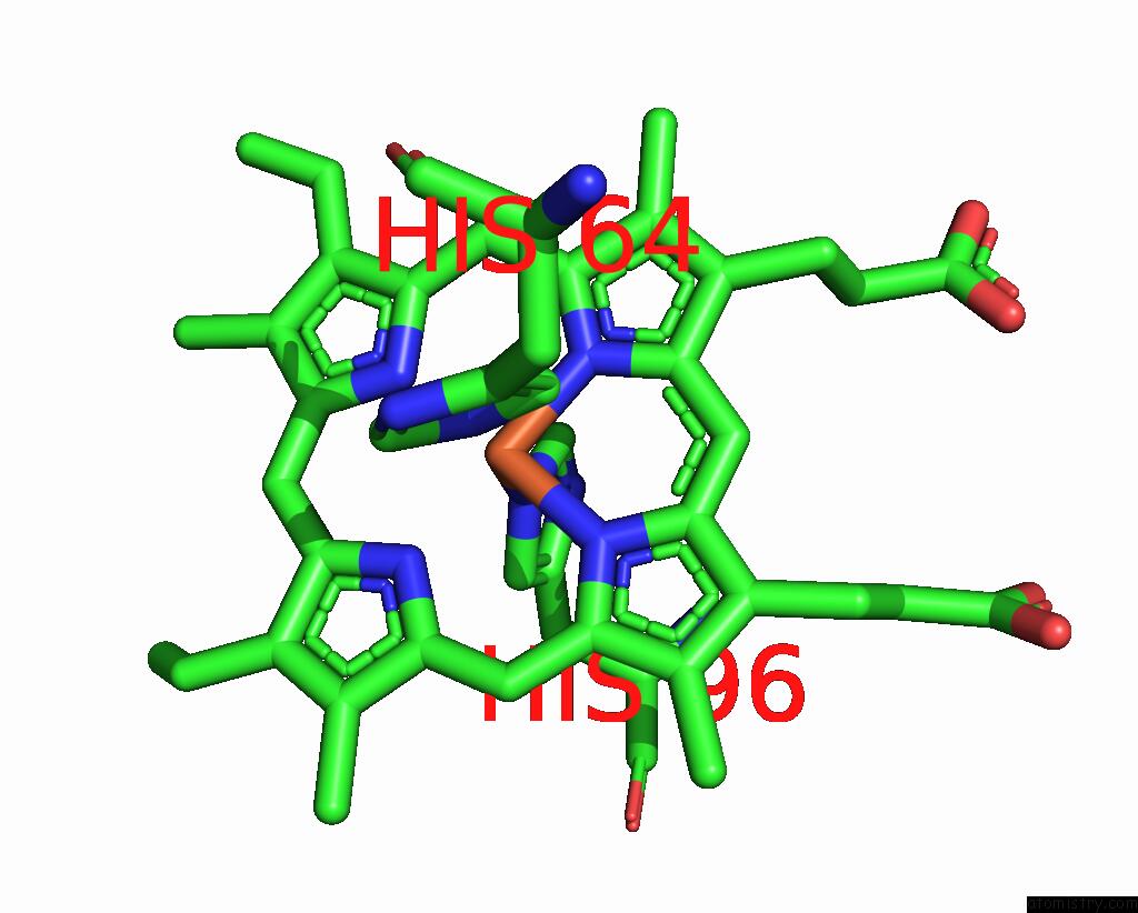
Mono view
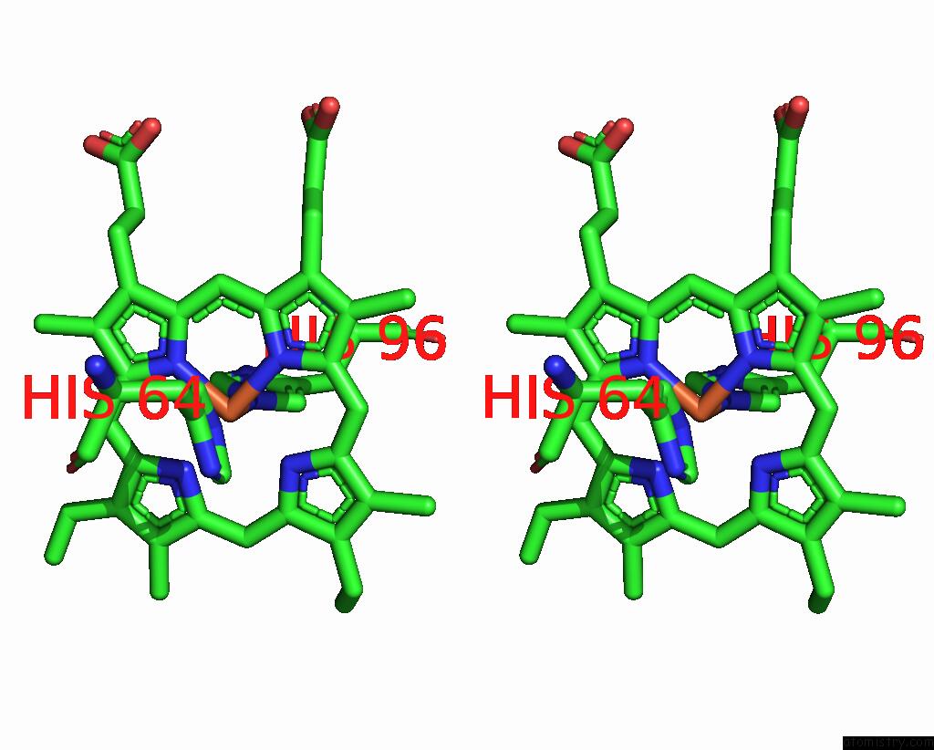
Stereo pair view
Mono view
Stereo pair view
A full contact list of Iron with other atoms in the Fe binding
site number 2 of The X-Ray Structure of Human Neuroglobin C120S Mutant within 5.0Å range:
|
Iron binding site 3 out of 3 in 8wub
Go back to
Iron binding site 3 out
of 3 in the The X-Ray Structure of Human Neuroglobin C120S Mutant
Mono view
Stereo pair view
Mono view
Stereo pair view
A full contact list of Iron with other atoms in the Fe binding
site number 3 of The X-Ray Structure of Human Neuroglobin C120S Mutant within 5.0Å range:
|
Reference:
Y.W.Lin,
H.Yuan.
The X-Ray Structure of Human Neuroglobin C120S Mutant To Be Published.
Page generated: Fri Aug 8 00:43:29 2025
Last articles
Fe in 9CQWFe in 9CQV
Fe in 9CQU
Fe in 9CQT
Fe in 9CQS
Fe in 9CJF
Fe in 9CQR
Fe in 9CQQ
Fe in 9CQP
Fe in 9CQO