Iron »
PDB 8wgh-8xcg »
8x9d »
Iron in PDB 8x9d: Crystal Structure of Co Dehydrogenase Mutant with Increased Affinity For Electron Mediators in High Peg Concentration
Enzymatic activity of Crystal Structure of Co Dehydrogenase Mutant with Increased Affinity For Electron Mediators in High Peg Concentration
All present enzymatic activity of Crystal Structure of Co Dehydrogenase Mutant with Increased Affinity For Electron Mediators in High Peg Concentration:
1.2.7.4;
1.2.7.4;
Protein crystallography data
The structure of Crystal Structure of Co Dehydrogenase Mutant with Increased Affinity For Electron Mediators in High Peg Concentration, PDB code: 8x9d
was solved by
H.H.Lee,
Y.Heo,
H.J.Yoon,
S.M.Kim,
S.Y.Kong,
with X-Ray Crystallography technique. A brief refinement statistics is given in the table below:
| Resolution Low / High (Å) | 33.02 / 2.11 |
| Space group | C 1 2 1 |
| Cell size a, b, c (Å), α, β, γ (°) | 112.045, 74.552, 71.309, 90, 111.53, 90 |
| R / Rfree (%) | 16.5 / 21.9 |
Other elements in 8x9d:
The structure of Crystal Structure of Co Dehydrogenase Mutant with Increased Affinity For Electron Mediators in High Peg Concentration also contains other interesting chemical elements:
| Nickel | (Ni) | 1 atom |
Iron Binding Sites:
Pages:
>>> Page 1 <<< Page 2, Binding sites: 11 - 11;Binding sites:
The binding sites of Iron atom in the Crystal Structure of Co Dehydrogenase Mutant with Increased Affinity For Electron Mediators in High Peg Concentration (pdb code 8x9d). This binding sites where shown within 5.0 Angstroms radius around Iron atom.In total 11 binding sites of Iron where determined in the Crystal Structure of Co Dehydrogenase Mutant with Increased Affinity For Electron Mediators in High Peg Concentration, PDB code: 8x9d:
Jump to Iron binding site number: 1; 2; 3; 4; 5; 6; 7; 8; 9; 10;
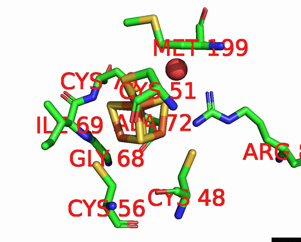
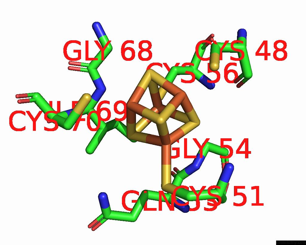
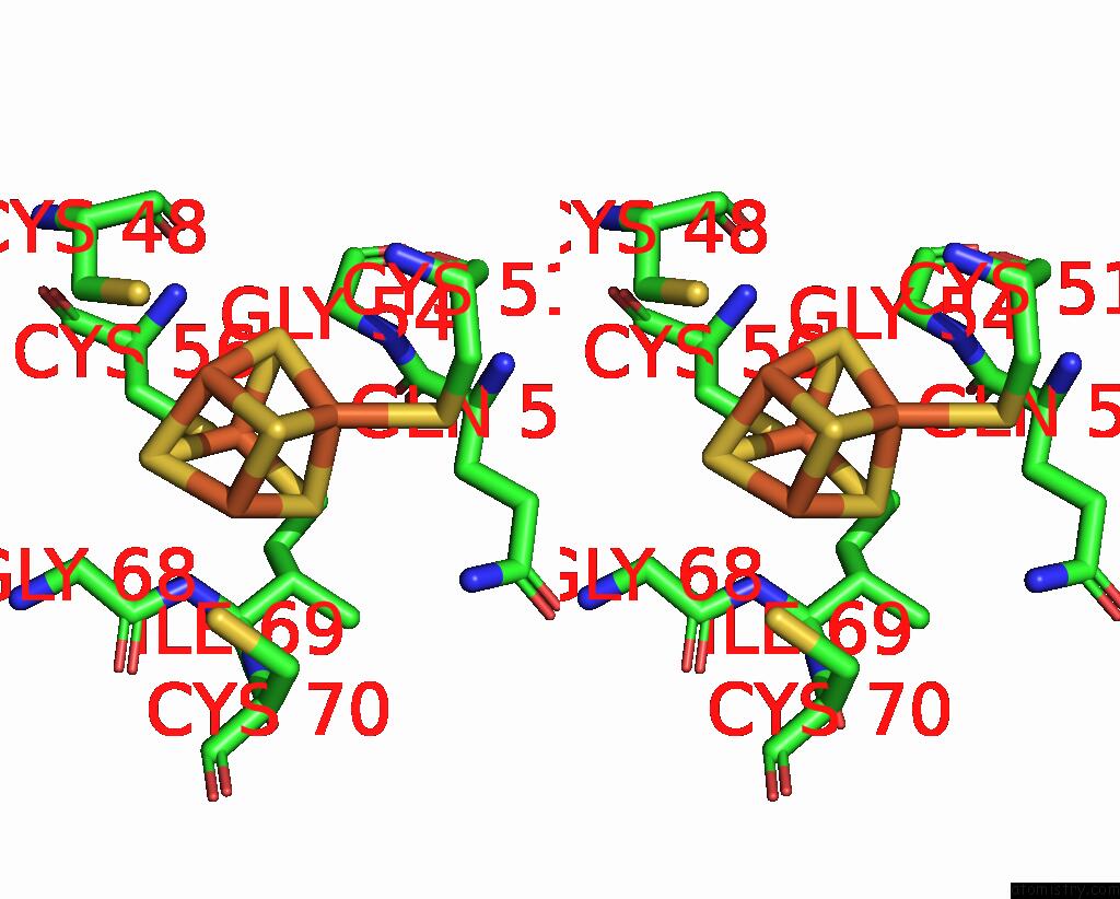
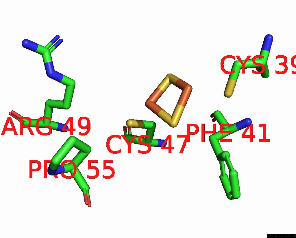
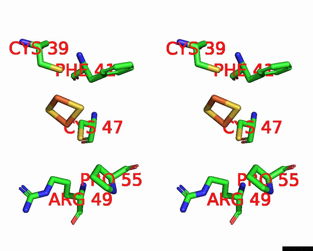
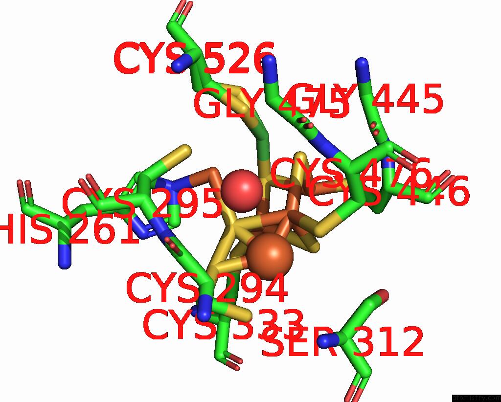
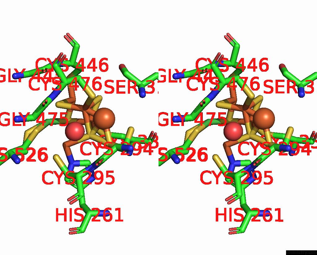
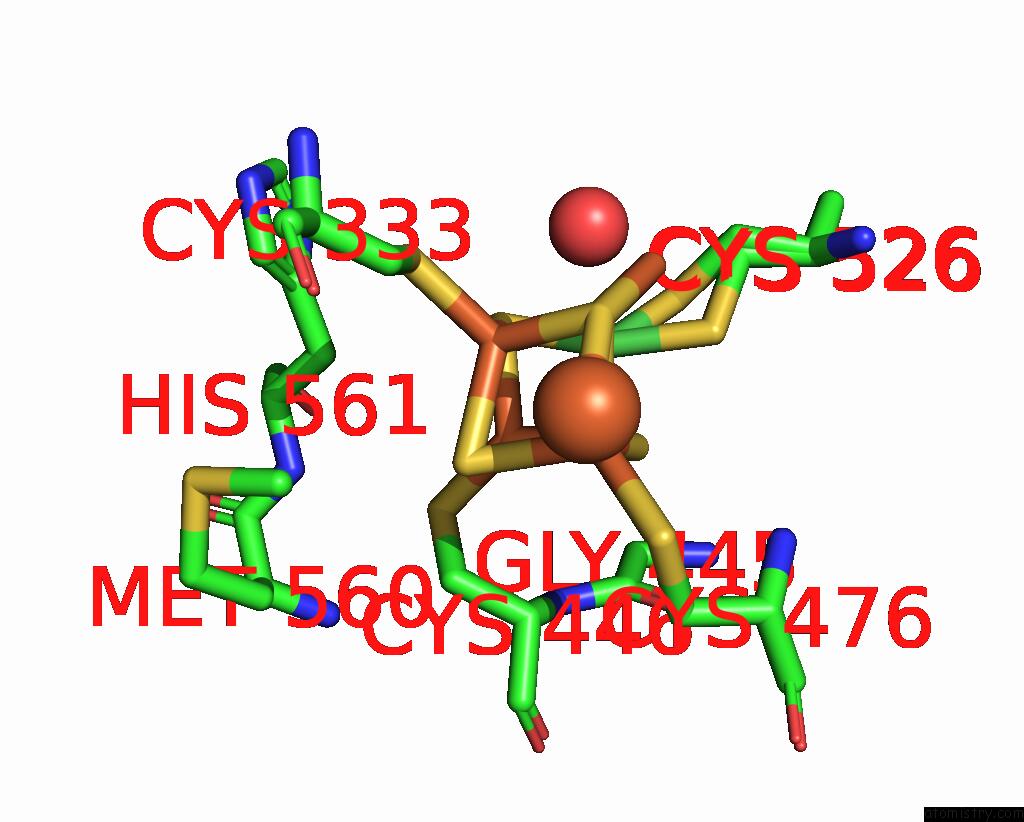
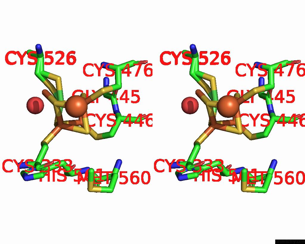
Iron binding site 1 out of 11 in 8x9d
Go back to
Iron binding site 1 out
of 11 in the Crystal Structure of Co Dehydrogenase Mutant with Increased Affinity For Electron Mediators in High Peg Concentration
Mono view
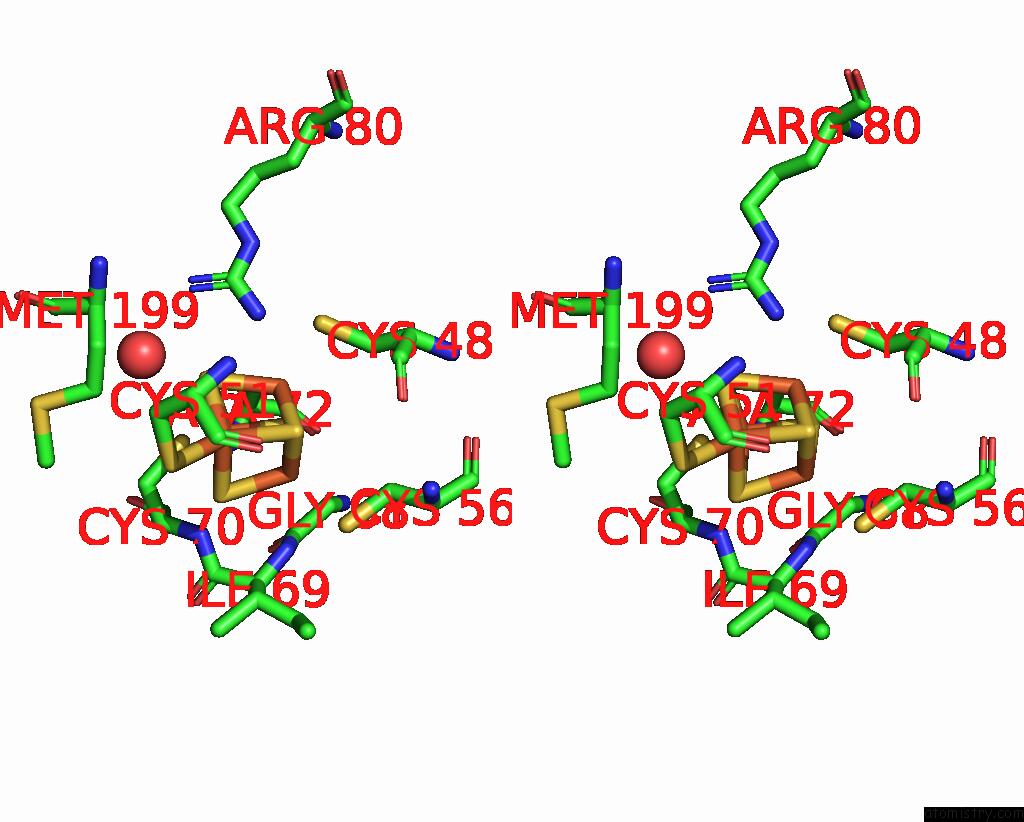
Stereo pair view
Mono view
Stereo pair view
A full contact list of Iron with other atoms in the Fe binding
site number 1 of Crystal Structure of Co Dehydrogenase Mutant with Increased Affinity For Electron Mediators in High Peg Concentration within 5.0Å range:
|
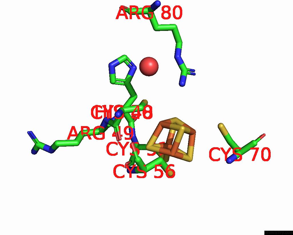
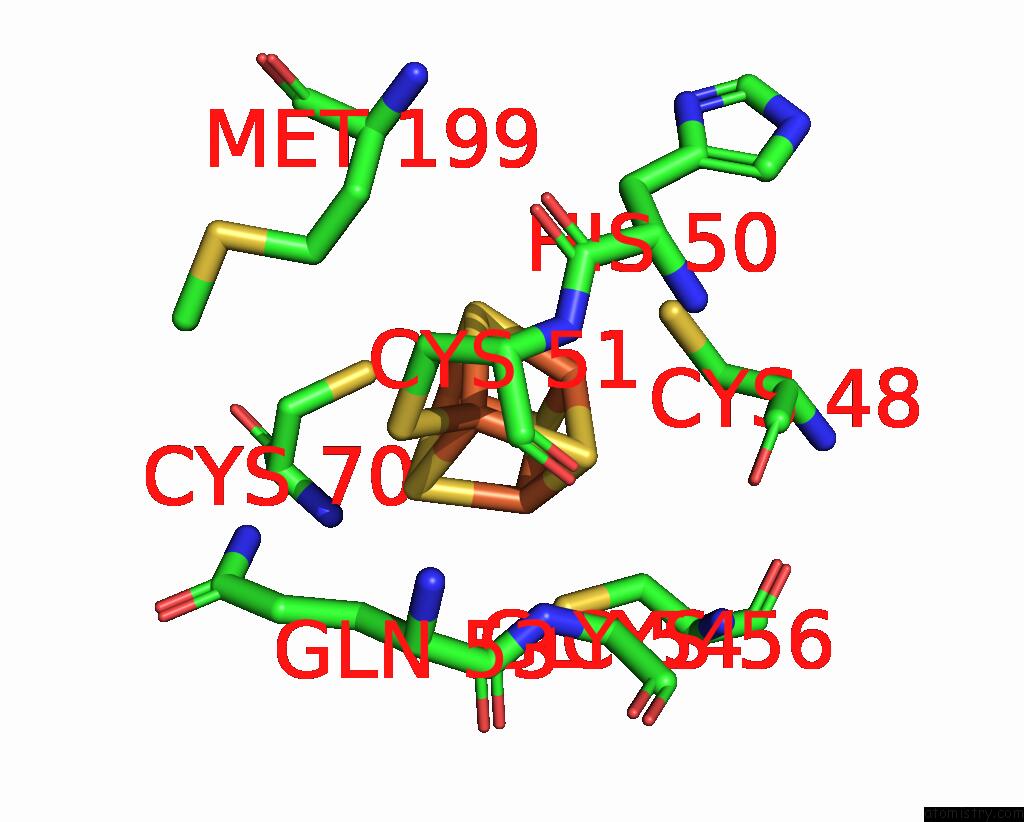
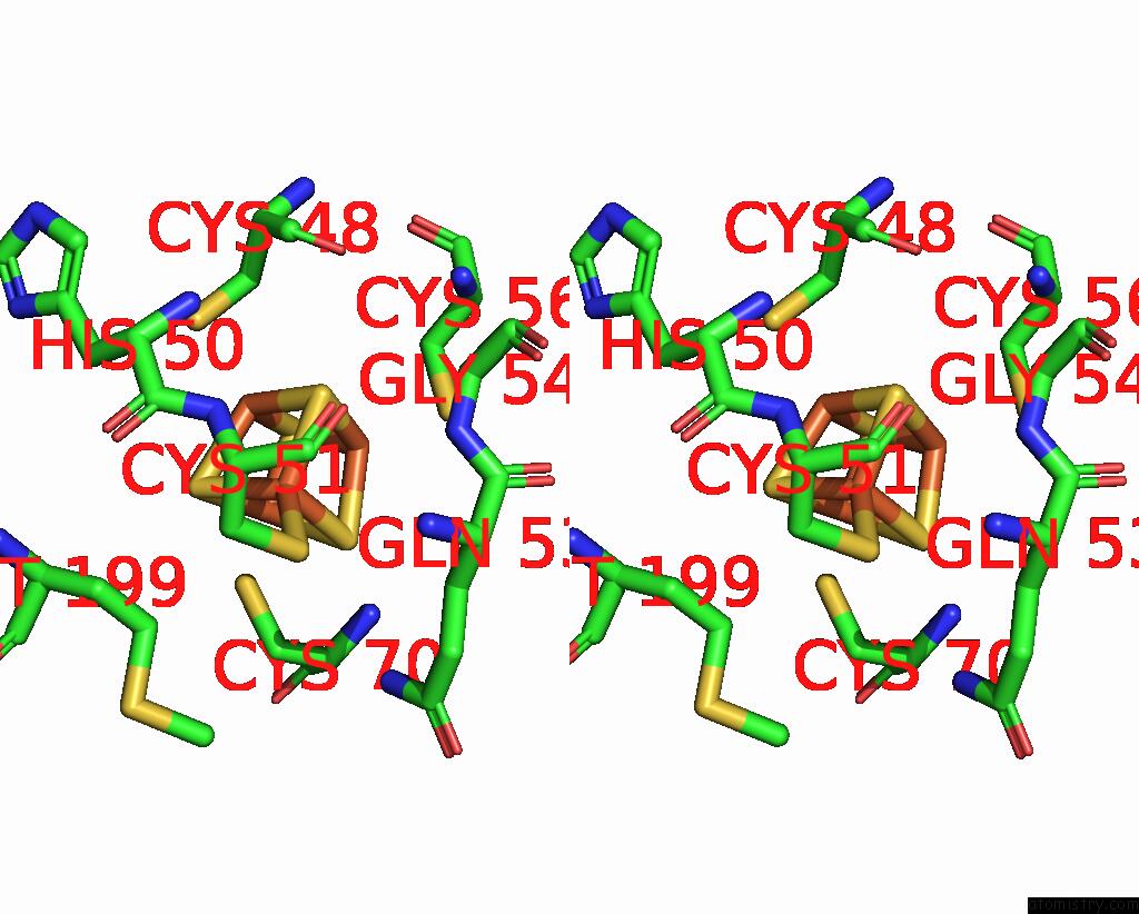
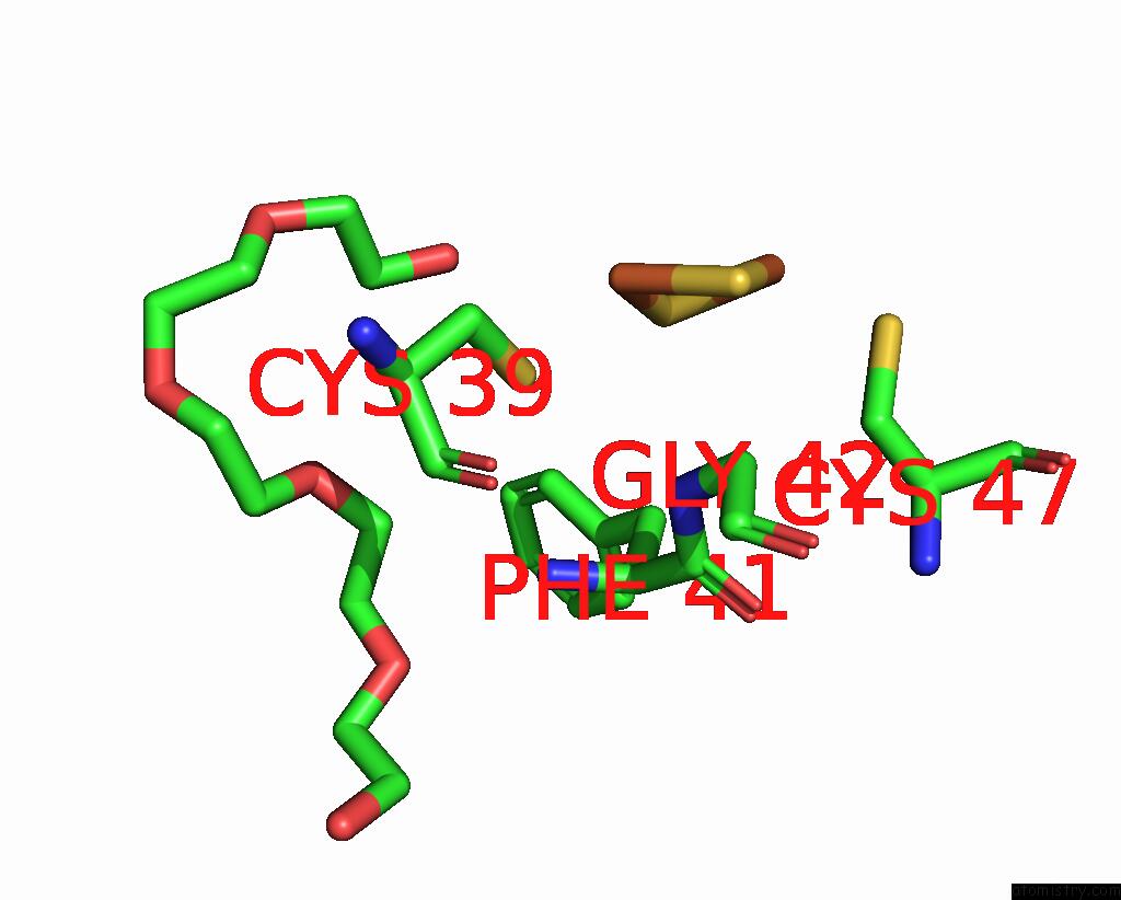
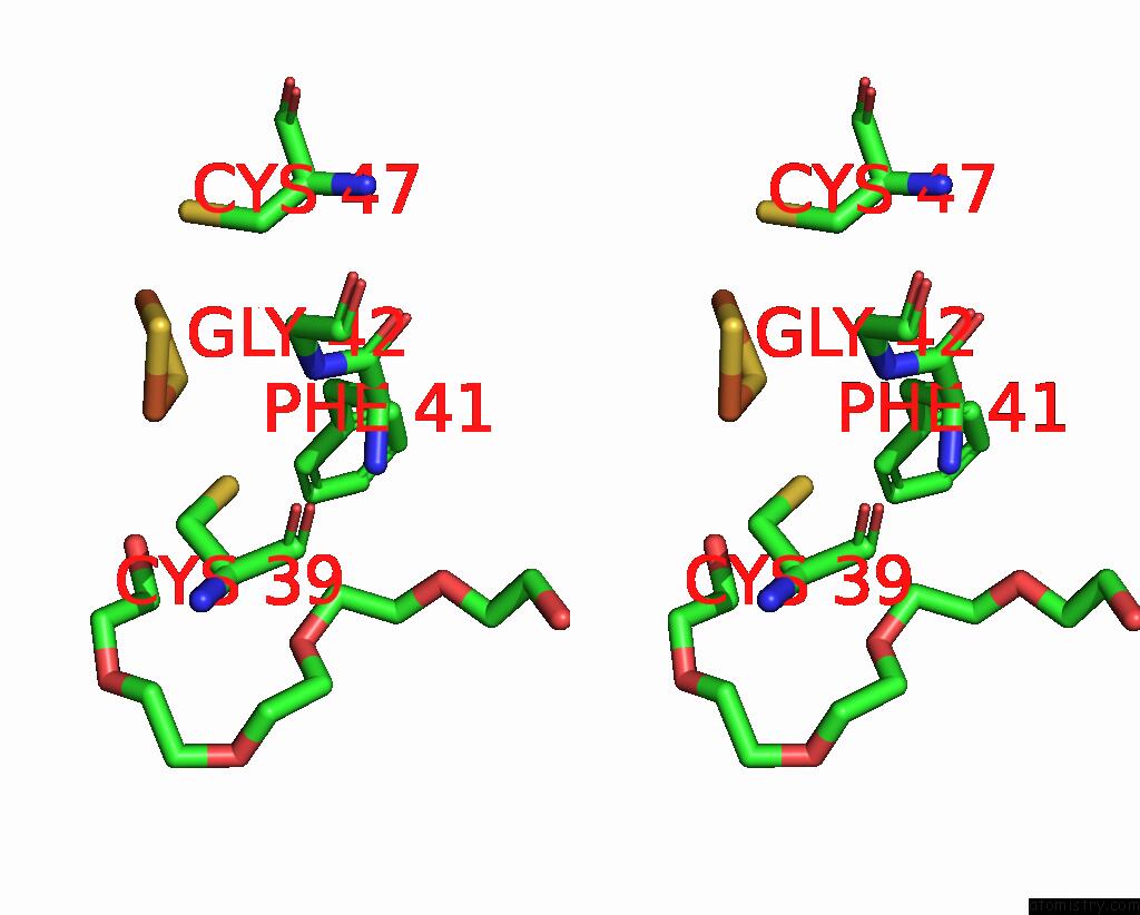
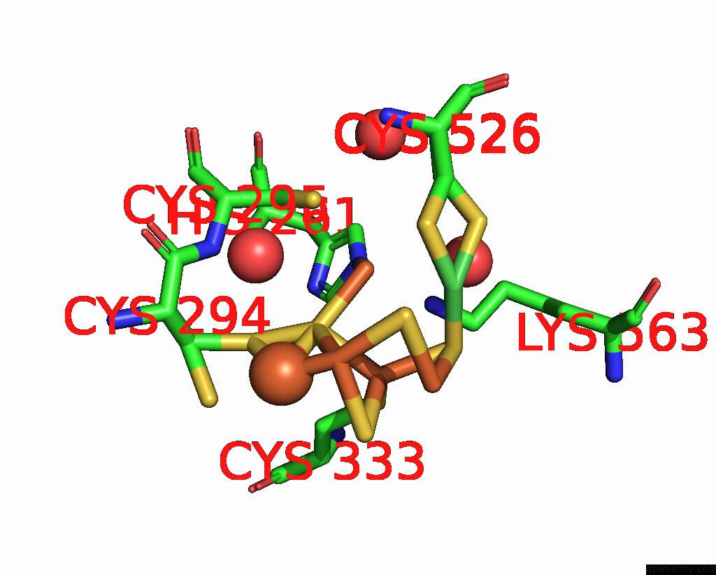
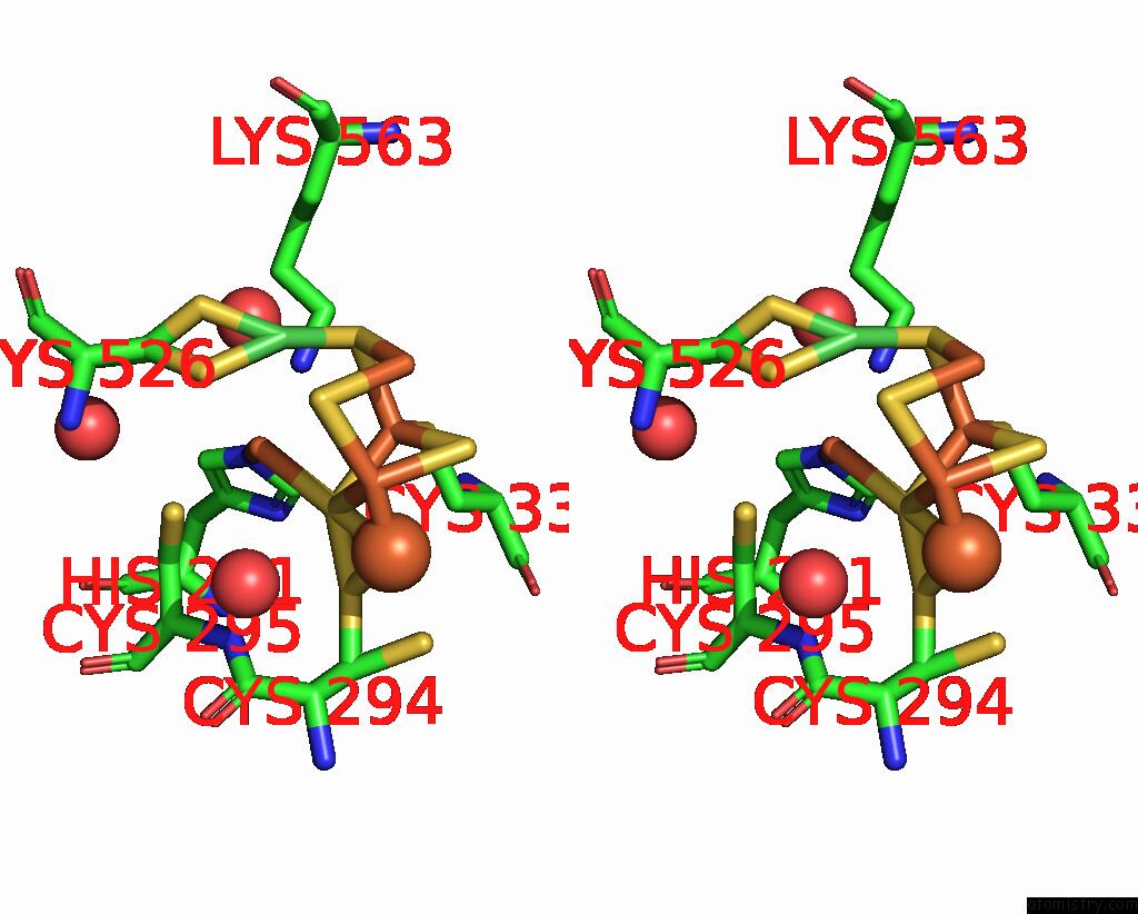
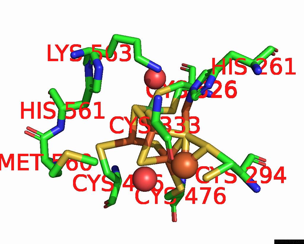
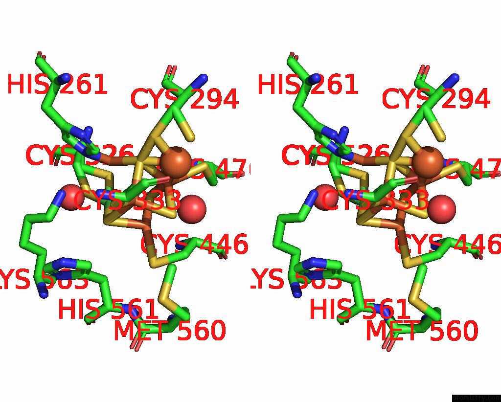
Iron binding site 2 out of 11 in 8x9d
Go back to
Iron binding site 2 out
of 11 in the Crystal Structure of Co Dehydrogenase Mutant with Increased Affinity For Electron Mediators in High Peg Concentration
Mono view
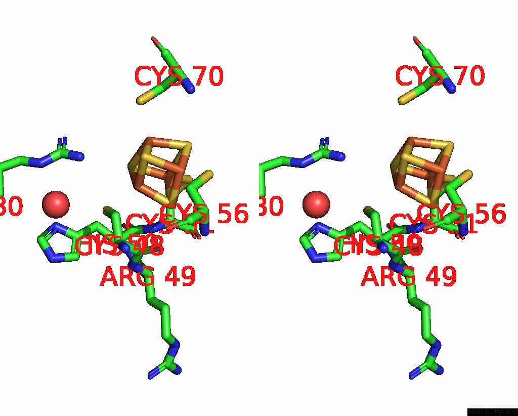
Stereo pair view
Mono view
Stereo pair view
A full contact list of Iron with other atoms in the Fe binding
site number 2 of Crystal Structure of Co Dehydrogenase Mutant with Increased Affinity For Electron Mediators in High Peg Concentration within 5.0Å range:
|
Iron binding site 3 out of 11 in 8x9d
Go back to
Iron binding site 3 out
of 11 in the Crystal Structure of Co Dehydrogenase Mutant with Increased Affinity For Electron Mediators in High Peg Concentration
Mono view
Stereo pair view
Mono view
Stereo pair view
A full contact list of Iron with other atoms in the Fe binding
site number 3 of Crystal Structure of Co Dehydrogenase Mutant with Increased Affinity For Electron Mediators in High Peg Concentration within 5.0Å range:
|
Iron binding site 4 out of 11 in 8x9d
Go back to
Iron binding site 4 out
of 11 in the Crystal Structure of Co Dehydrogenase Mutant with Increased Affinity For Electron Mediators in High Peg Concentration
Mono view
Stereo pair view
Mono view
Stereo pair view
A full contact list of Iron with other atoms in the Fe binding
site number 4 of Crystal Structure of Co Dehydrogenase Mutant with Increased Affinity For Electron Mediators in High Peg Concentration within 5.0Å range:
|
Iron binding site 5 out of 11 in 8x9d
Go back to
Iron binding site 5 out
of 11 in the Crystal Structure of Co Dehydrogenase Mutant with Increased Affinity For Electron Mediators in High Peg Concentration
Mono view
Stereo pair view
Mono view
Stereo pair view
A full contact list of Iron with other atoms in the Fe binding
site number 5 of Crystal Structure of Co Dehydrogenase Mutant with Increased Affinity For Electron Mediators in High Peg Concentration within 5.0Å range:
|
Iron binding site 6 out of 11 in 8x9d
Go back to
Iron binding site 6 out
of 11 in the Crystal Structure of Co Dehydrogenase Mutant with Increased Affinity For Electron Mediators in High Peg Concentration
Mono view
Stereo pair view
Mono view
Stereo pair view
A full contact list of Iron with other atoms in the Fe binding
site number 6 of Crystal Structure of Co Dehydrogenase Mutant with Increased Affinity For Electron Mediators in High Peg Concentration within 5.0Å range:
|
Iron binding site 7 out of 11 in 8x9d
Go back to
Iron binding site 7 out
of 11 in the Crystal Structure of Co Dehydrogenase Mutant with Increased Affinity For Electron Mediators in High Peg Concentration
Mono view
Stereo pair view
Mono view
Stereo pair view
A full contact list of Iron with other atoms in the Fe binding
site number 7 of Crystal Structure of Co Dehydrogenase Mutant with Increased Affinity For Electron Mediators in High Peg Concentration within 5.0Å range:
|
Iron binding site 8 out of 11 in 8x9d
Go back to
Iron binding site 8 out
of 11 in the Crystal Structure of Co Dehydrogenase Mutant with Increased Affinity For Electron Mediators in High Peg Concentration
Mono view
Stereo pair view
Mono view
Stereo pair view
A full contact list of Iron with other atoms in the Fe binding
site number 8 of Crystal Structure of Co Dehydrogenase Mutant with Increased Affinity For Electron Mediators in High Peg Concentration within 5.0Å range:
|
Iron binding site 9 out of 11 in 8x9d
Go back to
Iron binding site 9 out
of 11 in the Crystal Structure of Co Dehydrogenase Mutant with Increased Affinity For Electron Mediators in High Peg Concentration
Mono view
Stereo pair view
Mono view
Stereo pair view
A full contact list of Iron with other atoms in the Fe binding
site number 9 of Crystal Structure of Co Dehydrogenase Mutant with Increased Affinity For Electron Mediators in High Peg Concentration within 5.0Å range:
|
Iron binding site 10 out of 11 in 8x9d
Go back to
Iron binding site 10 out
of 11 in the Crystal Structure of Co Dehydrogenase Mutant with Increased Affinity For Electron Mediators in High Peg Concentration
Mono view
Stereo pair view
Mono view
Stereo pair view
A full contact list of Iron with other atoms in the Fe binding
site number 10 of Crystal Structure of Co Dehydrogenase Mutant with Increased Affinity For Electron Mediators in High Peg Concentration within 5.0Å range:
|
Reference:
S.M.Kim,
S.H.Kang,
J.Lee,
Y.Heo,
E.G.Poloniataki,
J.Kang,
H.J.Yoon,
S.Y.Kong,
Y.Yun,
H.Kim,
J.Ryu,
H.H.Lee,
Y.H.Kim.
Identifying A Key Spot For Electron Mediator-Interaction to Tailor Co Dehydrogenase'S Affinity Nat Commun V. 15 2732 2024.
ISSN: ESSN 2041-1723
DOI: 10.1038/S41467-024-46909-1
Page generated: Fri Aug 8 00:46:43 2025
ISSN: ESSN 2041-1723
DOI: 10.1038/S41467-024-46909-1
Last articles
Fe in 9CQWFe in 9CQV
Fe in 9CQU
Fe in 9CQT
Fe in 9CQS
Fe in 9CJF
Fe in 9CQR
Fe in 9CQQ
Fe in 9CQP
Fe in 9CQO