Iron in PDB 9ist: Crystal Structure of Cytochrome P450BM3 VI-18A12 Mutant Heme Domain with N-Decanoyl-L-Homoserine Lactone
Enzymatic activity of Crystal Structure of Cytochrome P450BM3 VI-18A12 Mutant Heme Domain with N-Decanoyl-L-Homoserine Lactone
All present enzymatic activity of Crystal Structure of Cytochrome P450BM3 VI-18A12 Mutant Heme Domain with N-Decanoyl-L-Homoserine Lactone:
1.14.14.1; 1.6.2.4;
1.14.14.1; 1.6.2.4;
Protein crystallography data
The structure of Crystal Structure of Cytochrome P450BM3 VI-18A12 Mutant Heme Domain with N-Decanoyl-L-Homoserine Lactone, PDB code: 9ist
was solved by
Y.Yokoyama,
H.Sugimoto,
O.Shoji,
with X-Ray Crystallography technique. A brief refinement statistics is given in the table below:
| Resolution Low / High (Å) | 47.00 / 2.27 |
| Space group | P 1 21 1 |
| Cell size a, b, c (Å), α, β, γ (°) | 58.482, 148.785, 61.374, 90, 98.91, 90 |
| R / Rfree (%) | 18.9 / 25.9 |
Iron Binding Sites:
The binding sites of Iron atom in the Crystal Structure of Cytochrome P450BM3 VI-18A12 Mutant Heme Domain with N-Decanoyl-L-Homoserine Lactone
(pdb code 9ist). This binding sites where shown within
5.0 Angstroms radius around Iron atom.
In total 2 binding sites of Iron where determined in the Crystal Structure of Cytochrome P450BM3 VI-18A12 Mutant Heme Domain with N-Decanoyl-L-Homoserine Lactone, PDB code: 9ist:
Jump to Iron binding site number: 1; 2;
In total 2 binding sites of Iron where determined in the Crystal Structure of Cytochrome P450BM3 VI-18A12 Mutant Heme Domain with N-Decanoyl-L-Homoserine Lactone, PDB code: 9ist:
Jump to Iron binding site number: 1; 2;
Iron binding site 1 out of 2 in 9ist
Go back to
Iron binding site 1 out
of 2 in the Crystal Structure of Cytochrome P450BM3 VI-18A12 Mutant Heme Domain with N-Decanoyl-L-Homoserine Lactone
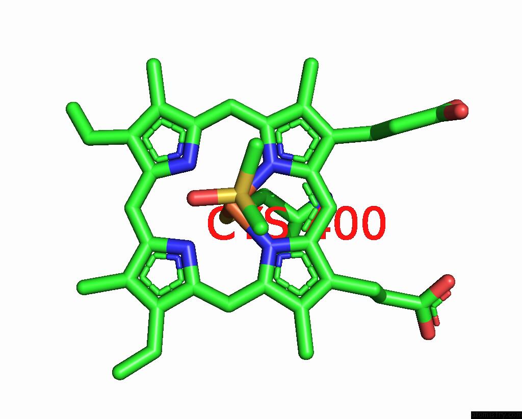
Mono view
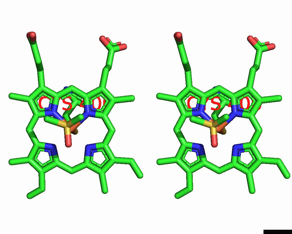
Stereo pair view
Mono view
Stereo pair view
A full contact list of Iron with other atoms in the Fe binding
site number 1 of Crystal Structure of Cytochrome P450BM3 VI-18A12 Mutant Heme Domain with N-Decanoyl-L-Homoserine Lactone within 5.0Å range:
|
Iron binding site 2 out of 2 in 9ist
Go back to
Iron binding site 2 out
of 2 in the Crystal Structure of Cytochrome P450BM3 VI-18A12 Mutant Heme Domain with N-Decanoyl-L-Homoserine Lactone
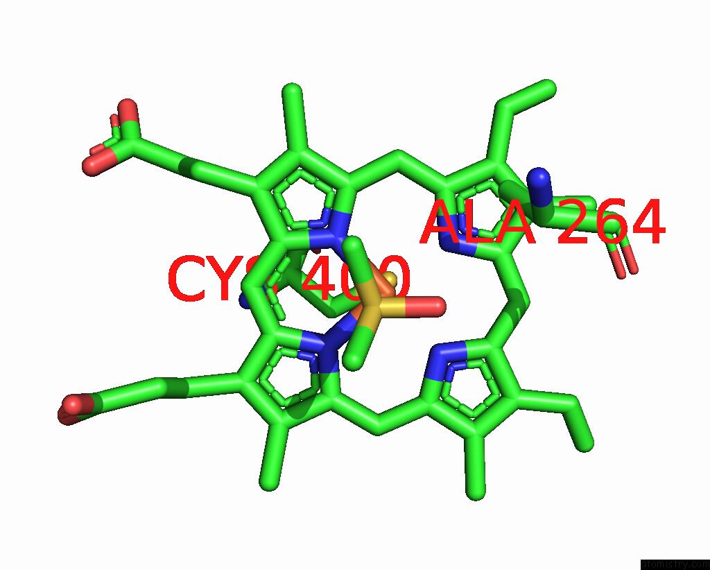
Mono view
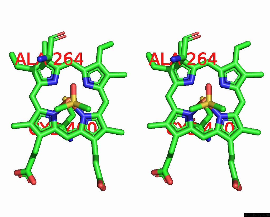
Stereo pair view
Mono view
Stereo pair view
A full contact list of Iron with other atoms in the Fe binding
site number 2 of Crystal Structure of Cytochrome P450BM3 VI-18A12 Mutant Heme Domain with N-Decanoyl-L-Homoserine Lactone within 5.0Å range:
|
Reference:
Y.Yokoyama,
S.Ariyasu,
M.Karasawa,
C.Kasai,
Y.Aiba,
H.Sugimoto,
O.Shoji.
Bacterial Acyl Homoserine Lactones Triggered Non-Native Substrate Hydroxylation Catalyzed By Directed-Evolution-Derived Cytochrome P450BM3 Mutants Chemcatchem 2024.
ISSN: ESSN 1867-3899
DOI: 10.1002/CCTC.202401641
Page generated: Tue Dec 10 20:12:40 2024
ISSN: ESSN 1867-3899
DOI: 10.1002/CCTC.202401641
Last articles
Zn in 9MJ5Zn in 9HNW
Zn in 9G0L
Zn in 9FNE
Zn in 9DZN
Zn in 9E0I
Zn in 9D32
Zn in 9DAK
Zn in 8ZXC
Zn in 8ZUF