Iron »
PDB 1a9w-1aop »
1aeu »
Iron in PDB 1aeu: Specificity of Ligand Binding in A Polar Cavity of Cytochrome C Peroxidase (2-Methylimidazole)
Enzymatic activity of Specificity of Ligand Binding in A Polar Cavity of Cytochrome C Peroxidase (2-Methylimidazole)
All present enzymatic activity of Specificity of Ligand Binding in A Polar Cavity of Cytochrome C Peroxidase (2-Methylimidazole):
1.11.1.5;
1.11.1.5;
Protein crystallography data
The structure of Specificity of Ligand Binding in A Polar Cavity of Cytochrome C Peroxidase (2-Methylimidazole), PDB code: 1aeu
was solved by
R.A.Musah,
G.M.Jensen,
M.M.Fitzgerald,
D.E.Mcree,
D.B.Goodin,
with X-Ray Crystallography technique. A brief refinement statistics is given in the table below:
| Resolution Low / High (Å) | 7.00 / 2.10 |
| Space group | P 21 21 21 |
| Cell size a, b, c (Å), α, β, γ (°) | 105.200, 74.300, 45.400, 90.00, 90.00, 90.00 |
| R / Rfree (%) | n/a / n/a |
Iron Binding Sites:
The binding sites of Iron atom in the Specificity of Ligand Binding in A Polar Cavity of Cytochrome C Peroxidase (2-Methylimidazole)
(pdb code 1aeu). This binding sites where shown within
5.0 Angstroms radius around Iron atom.
In total only one binding site of Iron was determined in the Specificity of Ligand Binding in A Polar Cavity of Cytochrome C Peroxidase (2-Methylimidazole), PDB code: 1aeu:
In total only one binding site of Iron was determined in the Specificity of Ligand Binding in A Polar Cavity of Cytochrome C Peroxidase (2-Methylimidazole), PDB code: 1aeu:
Iron binding site 1 out of 1 in 1aeu
Go back to
Iron binding site 1 out
of 1 in the Specificity of Ligand Binding in A Polar Cavity of Cytochrome C Peroxidase (2-Methylimidazole)
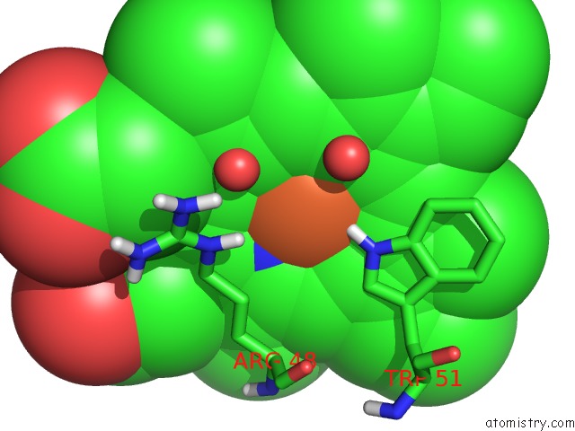
Mono view
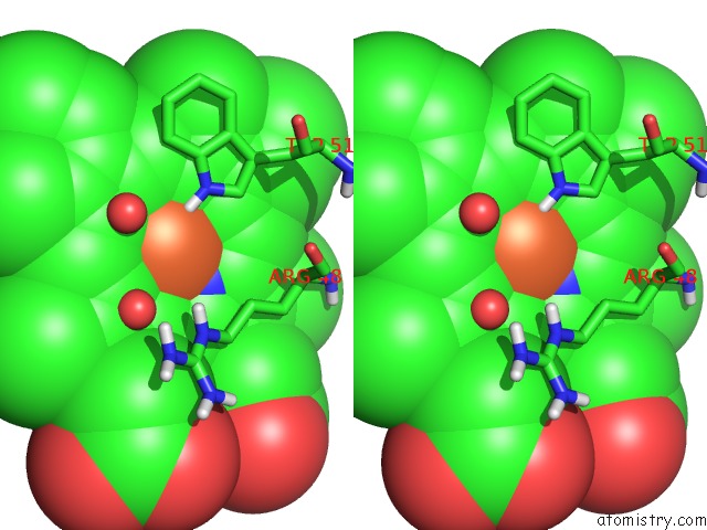
Stereo pair view
Mono view
Stereo pair view
A full contact list of Iron with other atoms in the Fe binding
site number 1 of Specificity of Ligand Binding in A Polar Cavity of Cytochrome C Peroxidase (2-Methylimidazole) within 5.0Å range:
|
Reference:
M.M.Fitzgerald,
R.A.Musah,
D.E.Mcree,
D.B.Goodin.
A Ligand-Gated, Hinged Loop Rearrangement Opens A Channel to A Buried Artificial Protein Cavity. Nat.Struct.Biol. V. 3 626 1996.
ISSN: ISSN 1072-8368
PubMed: 8673607
DOI: 10.1038/NSB0796-626
Page generated: Wed Jul 16 12:08:19 2025
ISSN: ISSN 1072-8368
PubMed: 8673607
DOI: 10.1038/NSB0796-626
Last articles
Mg in 4B2QMg in 4B3A
Mg in 4B1Z
Mg in 4B2P
Mg in 4B2M
Mg in 4B2K
Mg in 4B2J
Mg in 4B2D
Mg in 4B20
Mg in 4B2H