Iron »
PDB 1b2o-1biy »
1b2o »
Iron in PDB 1b2o: Clostridium Pasteurianum Rubredoxin G10VG43A Mutant
Protein crystallography data
The structure of Clostridium Pasteurianum Rubredoxin G10VG43A Mutant, PDB code: 1b2o
was solved by
M.J.Maher,
J.M.Guss,
M.C.J.Wilce,
A.G.Wedd,
with X-Ray Crystallography technique. A brief refinement statistics is given in the table below:
| Resolution Low / High (Å) | 30.00 / 1.90 |
| Space group | P 43 21 2 |
| Cell size a, b, c (Å), α, β, γ (°) | 61.850, 61.850, 80.450, 90.00, 90.00, 90.00 |
| R / Rfree (%) | 19.4 / 23.7 |
Iron Binding Sites:
The binding sites of Iron atom in the Clostridium Pasteurianum Rubredoxin G10VG43A Mutant
(pdb code 1b2o). This binding sites where shown within
5.0 Angstroms radius around Iron atom.
In total 2 binding sites of Iron where determined in the Clostridium Pasteurianum Rubredoxin G10VG43A Mutant, PDB code: 1b2o:
Jump to Iron binding site number: 1; 2;
In total 2 binding sites of Iron where determined in the Clostridium Pasteurianum Rubredoxin G10VG43A Mutant, PDB code: 1b2o:
Jump to Iron binding site number: 1; 2;
Iron binding site 1 out of 2 in 1b2o
Go back to
Iron binding site 1 out
of 2 in the Clostridium Pasteurianum Rubredoxin G10VG43A Mutant
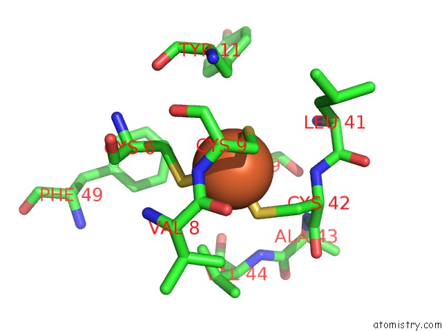
Mono view
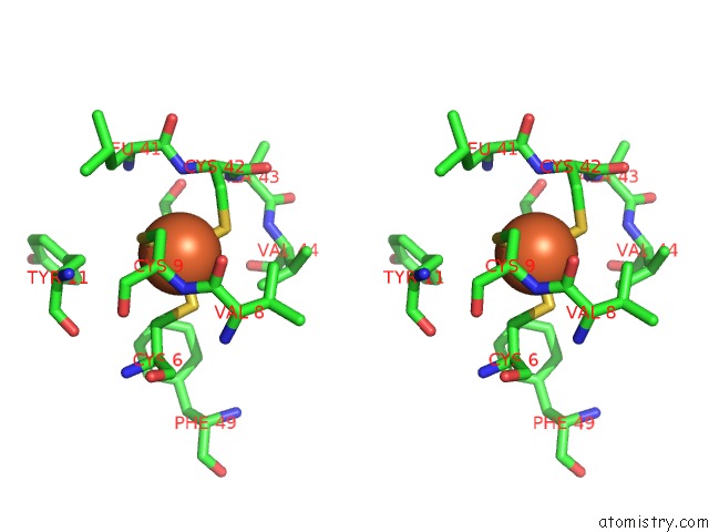
Stereo pair view
Mono view
Stereo pair view
A full contact list of Iron with other atoms in the Fe binding
site number 1 of Clostridium Pasteurianum Rubredoxin G10VG43A Mutant within 5.0Å range:
|
Iron binding site 2 out of 2 in 1b2o
Go back to
Iron binding site 2 out
of 2 in the Clostridium Pasteurianum Rubredoxin G10VG43A Mutant
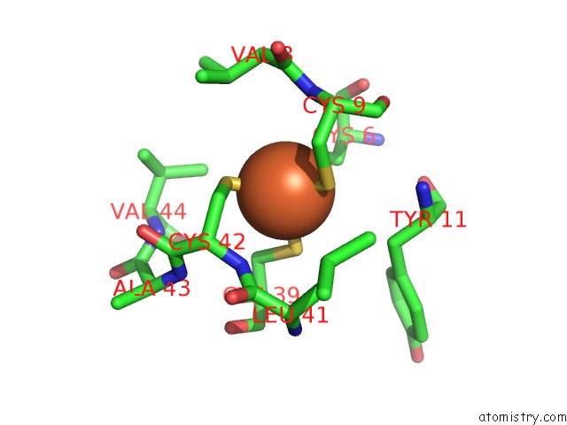
Mono view
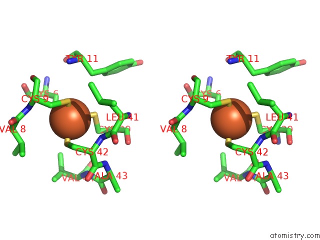
Stereo pair view
Mono view
Stereo pair view
A full contact list of Iron with other atoms in the Fe binding
site number 2 of Clostridium Pasteurianum Rubredoxin G10VG43A Mutant within 5.0Å range:
|
Reference:
M.J.Maher,
Z.Xiao,
M.C.Wilce,
J.M.Guss,
A.G.Wedd.
Rubredoxin From Clostridium Pasteurianum. Structures of G10A, G43A and G10VG43A Mutant Proteins. Mutation of Conserved Glycine 10 to Valine Causes the 9-10 Peptide Link to Invert. Acta Crystallogr.,Sect.D V. 55 962 1999.
ISSN: ISSN 0907-4449
PubMed: 10216292
DOI: 10.1107/S0907444999001900
Page generated: Wed Jul 16 12:28:01 2025
ISSN: ISSN 0907-4449
PubMed: 10216292
DOI: 10.1107/S0907444999001900
Last articles
Y in 2R3CY in 3BEJ
Y in 3BFW
Y in 2E81
Y in 2E80
Y in 2BEC
Y in 2C1C
Y in 1XVQ
Y in 2AHX
Y in 1XKG