Iron »
PDB 1b2o-1biy »
1b7v »
Iron in PDB 1b7v: Structure of the C-553 Cytochrome From Bacillus Pasteruii to 1.7 A Resolution
Protein crystallography data
The structure of Structure of the C-553 Cytochrome From Bacillus Pasteruii to 1.7 A Resolution, PDB code: 1b7v
was solved by
A.Gonzalez,
S.Benini,
W.R.Rypniewski,
K.S.Wilson,
S.Ciurli,
with X-Ray Crystallography technique. A brief refinement statistics is given in the table below:
| Resolution Low / High (Å) | 20.00 / 1.70 |
| Space group | P 21 21 21 |
| Cell size a, b, c (Å), α, β, γ (°) | 37.090, 39.160, 43.990, 90.00, 90.00, 90.00 |
| R / Rfree (%) | 17.4 / 20.6 |
Iron Binding Sites:
The binding sites of Iron atom in the Structure of the C-553 Cytochrome From Bacillus Pasteruii to 1.7 A Resolution
(pdb code 1b7v). This binding sites where shown within
5.0 Angstroms radius around Iron atom.
In total only one binding site of Iron was determined in the Structure of the C-553 Cytochrome From Bacillus Pasteruii to 1.7 A Resolution, PDB code: 1b7v:
In total only one binding site of Iron was determined in the Structure of the C-553 Cytochrome From Bacillus Pasteruii to 1.7 A Resolution, PDB code: 1b7v:
Iron binding site 1 out of 1 in 1b7v
Go back to
Iron binding site 1 out
of 1 in the Structure of the C-553 Cytochrome From Bacillus Pasteruii to 1.7 A Resolution
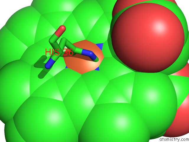
Mono view
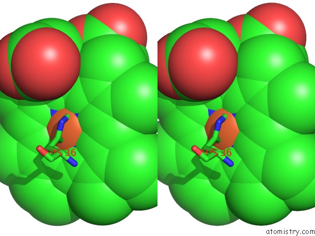
Stereo pair view
Mono view
Stereo pair view
A full contact list of Iron with other atoms in the Fe binding
site number 1 of Structure of the C-553 Cytochrome From Bacillus Pasteruii to 1.7 A Resolution within 5.0Å range:
|
Reference:
S.Benini,
A.Gonzalez,
W.R.Rypniewski,
K.S.Wilson,
J.J.Van Beeumen,
S.Ciurli.
Crystal Structure of Oxidized Bacillus Pasteurii Cytochrome C553 at 0.97-A Resolution. Biochemistry V. 39 13115 2000.
ISSN: ISSN 0006-2960
PubMed: 11052663
DOI: 10.1021/BI000402J
Page generated: Wed Jul 16 12:29:04 2025
ISSN: ISSN 0006-2960
PubMed: 11052663
DOI: 10.1021/BI000402J
Last articles
Y in 6MI5Y in 6THE
Y in 6NFQ
Y in 4WFD
Y in 4OR5
Y in 6KSV
Y in 6KSU
Y in 5U62
Y in 4YHU
Y in 5U5T