Iron »
PDB 1b2o-1biy »
1bbh »
Iron in PDB 1bbh: Atomic Structure of A Cytochrome C' with An Unusual Ligand-Controlled Dimer Dissociation at 1.8 Angstroms Resolution
Protein crystallography data
The structure of Atomic Structure of A Cytochrome C' with An Unusual Ligand-Controlled Dimer Dissociation at 1.8 Angstroms Resolution, PDB code: 1bbh
was solved by
Z.Ren,
D.E.Mcree,
with X-Ray Crystallography technique. A brief refinement statistics is given in the table below:
| Resolution Low / High (Å) | 5.00 / 1.80 |
| Space group | P 21 21 21 |
| Cell size a, b, c (Å), α, β, γ (°) | 49.200, 56.700, 98.800, 90.00, 90.00, 90.00 |
| R / Rfree (%) | 18.5 / n/a |
Iron Binding Sites:
The binding sites of Iron atom in the Atomic Structure of A Cytochrome C' with An Unusual Ligand-Controlled Dimer Dissociation at 1.8 Angstroms Resolution
(pdb code 1bbh). This binding sites where shown within
5.0 Angstroms radius around Iron atom.
In total 2 binding sites of Iron where determined in the Atomic Structure of A Cytochrome C' with An Unusual Ligand-Controlled Dimer Dissociation at 1.8 Angstroms Resolution, PDB code: 1bbh:
Jump to Iron binding site number: 1; 2;
In total 2 binding sites of Iron where determined in the Atomic Structure of A Cytochrome C' with An Unusual Ligand-Controlled Dimer Dissociation at 1.8 Angstroms Resolution, PDB code: 1bbh:
Jump to Iron binding site number: 1; 2;
Iron binding site 1 out of 2 in 1bbh
Go back to
Iron binding site 1 out
of 2 in the Atomic Structure of A Cytochrome C' with An Unusual Ligand-Controlled Dimer Dissociation at 1.8 Angstroms Resolution
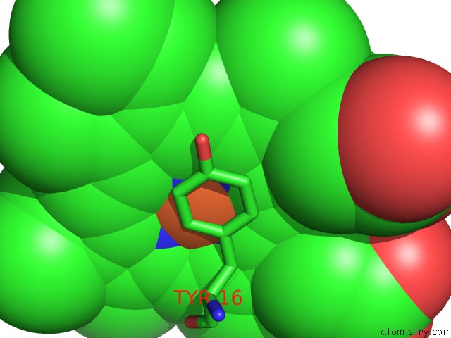
Mono view
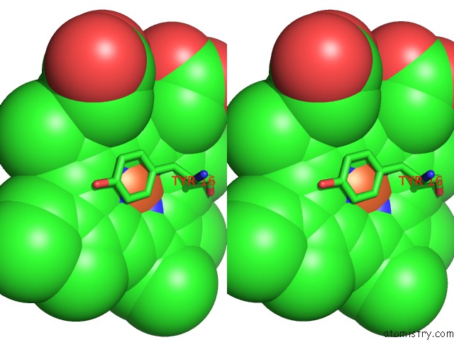
Stereo pair view
Mono view
Stereo pair view
A full contact list of Iron with other atoms in the Fe binding
site number 1 of Atomic Structure of A Cytochrome C' with An Unusual Ligand-Controlled Dimer Dissociation at 1.8 Angstroms Resolution within 5.0Å range:
|
Iron binding site 2 out of 2 in 1bbh
Go back to
Iron binding site 2 out
of 2 in the Atomic Structure of A Cytochrome C' with An Unusual Ligand-Controlled Dimer Dissociation at 1.8 Angstroms Resolution
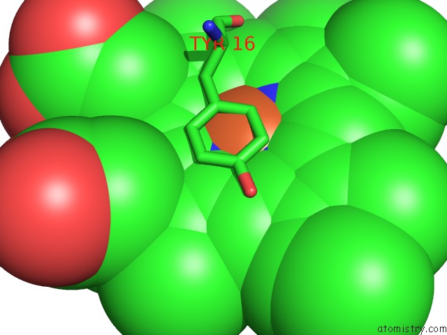
Mono view
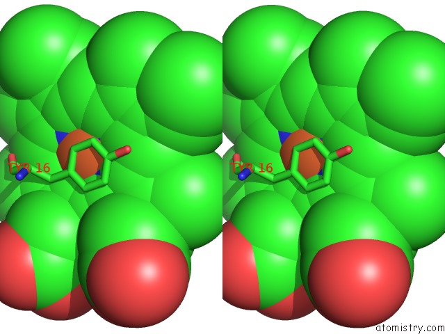
Stereo pair view
Mono view
Stereo pair view
A full contact list of Iron with other atoms in the Fe binding
site number 2 of Atomic Structure of A Cytochrome C' with An Unusual Ligand-Controlled Dimer Dissociation at 1.8 Angstroms Resolution within 5.0Å range:
|
Reference:
Z.Ren,
T.Meyer,
D.E.Mcree.
Atomic Structure of A Cytochrome C' with An Unusual Ligand-Controlled Dimer Dissociation at 1.8 A Resolution. J.Mol.Biol. V. 234 433 1993.
ISSN: ISSN 0022-2836
PubMed: 8230224
DOI: 10.1006/JMBI.1993.1597
Page generated: Wed Jul 16 12:31:18 2025
ISSN: ISSN 0022-2836
PubMed: 8230224
DOI: 10.1006/JMBI.1993.1597
Last articles
Y in 2R3CY in 3BEJ
Y in 3BFW
Y in 2E81
Y in 2E80
Y in 2BEC
Y in 2C1C
Y in 1XVQ
Y in 2AHX
Y in 1XKG