Iron »
PDB 1b2o-1biy »
1biy »
Iron in PDB 1biy: Structure of Diferric Buffalo Lactoferrin
Protein crystallography data
The structure of Structure of Diferric Buffalo Lactoferrin, PDB code: 1biy
was solved by
S.Karthikeyan,
S.Yadav,
T.P.Singh,
with X-Ray Crystallography technique. A brief refinement statistics is given in the table below:
| Resolution Low / High (Å) | 17.00 / 3.37 |
| Space group | P 1 21 1 |
| Cell size a, b, c (Å), α, β, γ (°) | 56.797, 101.436, 76.275, 90.00, 104.88, 90.00 |
| R / Rfree (%) | 21.8 / 31.8 |
Iron Binding Sites:
The binding sites of Iron atom in the Structure of Diferric Buffalo Lactoferrin
(pdb code 1biy). This binding sites where shown within
5.0 Angstroms radius around Iron atom.
In total 2 binding sites of Iron where determined in the Structure of Diferric Buffalo Lactoferrin, PDB code: 1biy:
Jump to Iron binding site number: 1; 2;
In total 2 binding sites of Iron where determined in the Structure of Diferric Buffalo Lactoferrin, PDB code: 1biy:
Jump to Iron binding site number: 1; 2;
Iron binding site 1 out of 2 in 1biy
Go back to
Iron binding site 1 out
of 2 in the Structure of Diferric Buffalo Lactoferrin
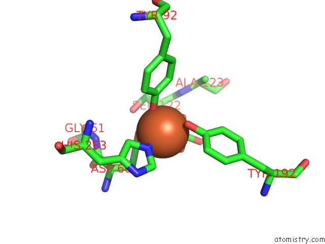
Mono view
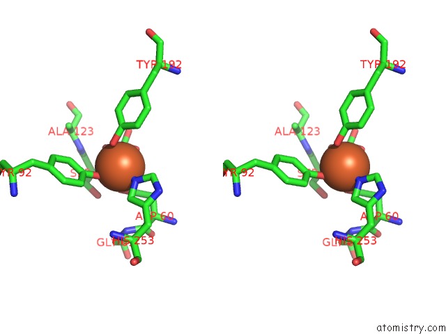
Stereo pair view
Mono view
Stereo pair view
A full contact list of Iron with other atoms in the Fe binding
site number 1 of Structure of Diferric Buffalo Lactoferrin within 5.0Å range:
|
Iron binding site 2 out of 2 in 1biy
Go back to
Iron binding site 2 out
of 2 in the Structure of Diferric Buffalo Lactoferrin
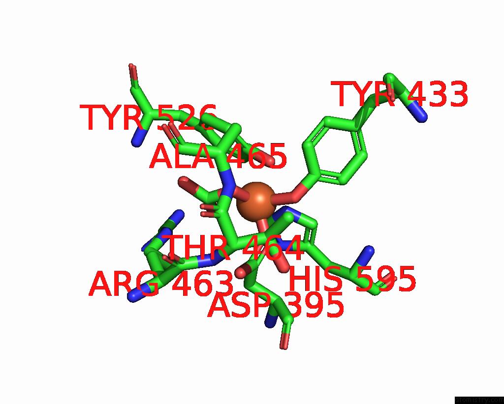
Mono view
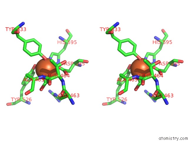
Stereo pair view
Mono view
Stereo pair view
A full contact list of Iron with other atoms in the Fe binding
site number 2 of Structure of Diferric Buffalo Lactoferrin within 5.0Å range:
|
Reference:
S.Karthikeyan,
S.Yadav,
M.Paramasivam,
A.Srinivasan,
T.P.Singh.
Structure of Buffalo Lactoferrin at 3.3 A Resolution at 277 K. Acta Crystallogr.,Sect.D V. 56 684 2000.
ISSN: ISSN 0907-4449
PubMed: 10818344
DOI: 10.1107/S0907444900005151
Page generated: Wed Jul 16 12:37:32 2025
ISSN: ISSN 0907-4449
PubMed: 10818344
DOI: 10.1107/S0907444900005151
Last articles
Y in 3SAHY in 3SAG
Y in 3SYX
Y in 3PH6
Y in 3SAF
Y in 3K38
Y in 3PH5
Y in 3N9D
Y in 3K3A
Y in 3K39