Iron »
PDB 1bj9-1c53 »
1bje »
Iron in PDB 1bje: H64T Variant of Myoglobin (Horse Heart) Recombinant Wild- Type Complexed with Azide
Protein crystallography data
The structure of H64T Variant of Myoglobin (Horse Heart) Recombinant Wild- Type Complexed with Azide, PDB code: 1bje
was solved by
R.Maurus,
G.D.Brayer,
with X-Ray Crystallography technique. A brief refinement statistics is given in the table below:
| Resolution Low / High (Å) | 6.00 / 1.80 |
| Space group | P 1 21 1 |
| Cell size a, b, c (Å), α, β, γ (°) | 64.019, 28.812, 35.840, 90.00, 106.95, 90.00 |
| R / Rfree (%) | 17.8 / n/a |
Iron Binding Sites:
The binding sites of Iron atom in the H64T Variant of Myoglobin (Horse Heart) Recombinant Wild- Type Complexed with Azide
(pdb code 1bje). This binding sites where shown within
5.0 Angstroms radius around Iron atom.
In total only one binding site of Iron was determined in the H64T Variant of Myoglobin (Horse Heart) Recombinant Wild- Type Complexed with Azide, PDB code: 1bje:
In total only one binding site of Iron was determined in the H64T Variant of Myoglobin (Horse Heart) Recombinant Wild- Type Complexed with Azide, PDB code: 1bje:
Iron binding site 1 out of 1 in 1bje
Go back to
Iron binding site 1 out
of 1 in the H64T Variant of Myoglobin (Horse Heart) Recombinant Wild- Type Complexed with Azide
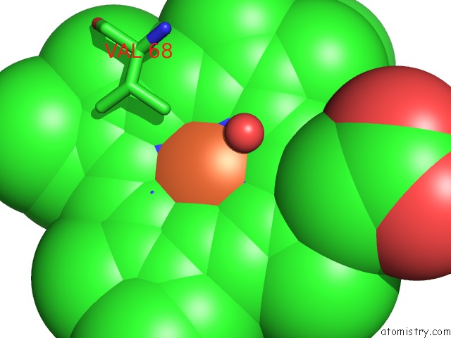
Mono view
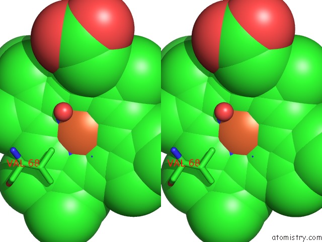
Stereo pair view
Mono view
Stereo pair view
A full contact list of Iron with other atoms in the Fe binding
site number 1 of H64T Variant of Myoglobin (Horse Heart) Recombinant Wild- Type Complexed with Azide within 5.0Å range:
|
Reference:
R.Maurus,
R.Bogumil,
N.T.Nguyen,
A.G.Mauk,
G.Brayer.
Structural and Spectroscopic Studies of Azide Complexes of Horse Heart Myoglobin and the His-64-->Thr Variant. Biochem.J. V. 332 67 1998.
ISSN: ISSN 0264-6021
PubMed: 9576852
Page generated: Wed Jul 16 12:38:44 2025
ISSN: ISSN 0264-6021
PubMed: 9576852
Last articles
Mg in 5P9RMg in 5P9T
Mg in 5P9S
Mg in 5P9P
Mg in 5P9O
Mg in 5P9N
Mg in 5P9E
Mg in 5P9D
Mg in 5P9C
Mg in 5P9B