Atomistry » Iron » PDB 1bj9-1c53 » 1bvb
Atomistry »
  Iron »
    PDB 1bj9-1c53 »
      1bvb »

Iron in PDB 1bvb: Heme-Packing Motifs Revealed By the Crystal Structure of Cytochrome C554 From Nitrosomonas Europaea

Protein crystallography data

The structure of Heme-Packing Motifs Revealed By the Crystal Structure of Cytochrome C554 From Nitrosomonas Europaea, PDB code: 1bvb was solved by T.M.Iverson, D.M.Arciero, B.T.Hsu, M.S.P.Logan, A.B.Hooper, D.C.Rees, with X-Ray Crystallography technique. A brief refinement statistics is given in the table below:

Resolution Low / High (Å) 20.00 / 2.60
Space group P 43 2 2
Cell size a, b, c (Å), α, β, γ (°) 68.200, 68.200, 168.833, 90.00, 90.00, 90.00
R / Rfree (%) 20.8 / 25.4

Iron Binding Sites:

The binding sites of Iron atom in the Heme-Packing Motifs Revealed By the Crystal Structure of Cytochrome C554 From Nitrosomonas Europaea (pdb code 1bvb). This binding sites where shown within 5.0 Angstroms radius around Iron atom.
In total 4 binding sites of Iron where determined in the Heme-Packing Motifs Revealed By the Crystal Structure of Cytochrome C554 From Nitrosomonas Europaea, PDB code: 1bvb:
Jump to Iron binding site number: 1; 2; 3; 4;

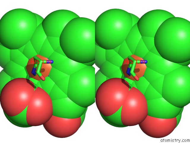
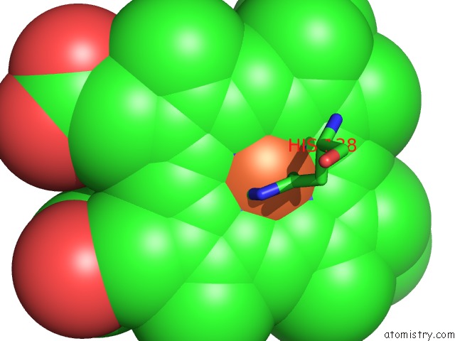
Iron binding site 1 out of 4 in 1bvb

Go back to Iron Binding Sites List in 1bvb
Iron binding site 1 out of 4 in the Heme-Packing Motifs Revealed By the Crystal Structure of Cytochrome C554 From Nitrosomonas Europaea


Mono view


Stereo pair view

A full contact list of Iron with other atoms in the Fe binding site number 1 of Heme-Packing Motifs Revealed By the Crystal Structure of Cytochrome C554 From Nitrosomonas Europaea within 5.0Å range:
probe atom residue distance (Å) B Occ
A:Fe213

b:15.1
occ:1.00
FE A:HEM213 0.0 15.1 1.0
NC A:HEM213 2.0 19.2 1.0
ND A:HEM213 2.0 20.3 1.0
NB A:HEM213 2.0 19.9 1.0
NA A:HEM213 2.0 19.1 1.0
NE2 A:HIS15 2.0 14.4 1.0
ND1 A:HIS102 2.2 22.9 1.0
C1C A:HEM213 3.0 16.6 1.0
C4C A:HEM213 3.0 18.0 1.0
C1A A:HEM213 3.0 21.2 1.0
C4B A:HEM213 3.0 21.4 1.0
CE1 A:HIS15 3.0 13.2 1.0
C4A A:HEM213 3.0 21.3 1.0
C4D A:HEM213 3.0 20.3 1.0
C1D A:HEM213 3.0 17.8 1.0
CE1 A:HIS102 3.0 25.0 1.0
C1B A:HEM213 3.1 17.8 1.0
CD2 A:HIS15 3.1 12.7 1.0
CG A:HIS102 3.3 29.9 1.0
CHC A:HEM213 3.4 20.0 1.0
CHA A:HEM213 3.4 19.5 1.0
CHD A:HEM213 3.4 14.7 1.0
CHB A:HEM213 3.4 20.8 1.0
CB A:HIS102 3.8 29.3 1.0
ND1 A:HIS15 4.2 16.0 1.0
C3C A:HEM213 4.2 19.0 1.0
CG A:HIS15 4.2 18.4 1.0
C2C A:HEM213 4.2 15.0 1.0
NE2 A:HIS102 4.3 27.3 1.0
C2A A:HEM213 4.3 20.4 1.0
C3D A:HEM213 4.3 19.5 1.0
C2D A:HEM213 4.3 19.3 1.0
C3A A:HEM213 4.3 23.1 1.0
C3B A:HEM213 4.3 18.1 1.0
C2B A:HEM213 4.3 17.7 1.0
CD2 A:HIS102 4.4 30.9 1.0

Iron binding site 2 out of 4 in 1bvb

Go back to Iron Binding Sites List in 1bvb
Iron binding site 2 out of 4 in the Heme-Packing Motifs Revealed By the Crystal Structure of Cytochrome C554 From Nitrosomonas Europaea


Mono view


Stereo pair view

A full contact list of Iron with other atoms in the Fe binding site number 2 of Heme-Packing Motifs Revealed By the Crystal Structure of Cytochrome C554 From Nitrosomonas Europaea within 5.0Å range:
probe atom residue distance (Å) B Occ
A:Fe214

b:13.4
occ:1.00
FE A:HEM214 0.0 13.4 1.0
ND A:HEM214 2.0 15.4 1.0
NC A:HEM214 2.0 8.9 1.0
NA A:HEM214 2.0 7.1 1.0
NB A:HEM214 2.0 10.8 1.0
NE2 A:HIS64 2.4 10.7 1.0
C1A A:HEM214 3.0 13.0 1.0
C1C A:HEM214 3.0 10.4 1.0
C4D A:HEM214 3.0 16.3 1.0
C4C A:HEM214 3.0 13.7 1.0
C1D A:HEM214 3.1 14.4 1.0
C4B A:HEM214 3.1 11.9 1.0
C4A A:HEM214 3.1 9.9 1.0
C1B A:HEM214 3.1 11.0 1.0
CE1 A:HIS64 3.2 15.5 1.0
CHA A:HEM214 3.4 11.4 1.0
CHC A:HEM214 3.4 8.9 1.0
CHD A:HEM214 3.4 12.8 1.0
CHB A:HEM214 3.5 11.3 1.0
CD2 A:HIS64 3.6 9.3 1.0
CD A:PRO155 4.1 17.6 1.0
CD2 A:PHE156 4.1 14.1 1.0
CE2 A:PHE156 4.2 9.4 1.0
C3C A:HEM214 4.3 8.6 1.0
C2D A:HEM214 4.3 16.1 1.0
C3D A:HEM214 4.3 15.4 1.0
C2C A:HEM214 4.3 12.8 1.0
C2A A:HEM214 4.3 10.5 1.0
C3B A:HEM214 4.3 9.5 1.0
C2B A:HEM214 4.3 6.1 1.0
C3A A:HEM214 4.4 9.8 1.0
ND1 A:HIS64 4.4 10.3 1.0
CG A:HIS64 4.7 9.9 1.0
CG A:PRO155 4.7 12.4 1.0
CMD A:HEM216 4.7 12.4 1.0

Iron binding site 3 out of 4 in 1bvb

Go back to Iron Binding Sites List in 1bvb
Iron binding site 3 out of 4 in the Heme-Packing Motifs Revealed By the Crystal Structure of Cytochrome C554 From Nitrosomonas Europaea


Mono view


Stereo pair view

A full contact list of Iron with other atoms in the Fe binding site number 3 of Heme-Packing Motifs Revealed By the Crystal Structure of Cytochrome C554 From Nitrosomonas Europaea within 5.0Å range:
probe atom residue distance (Å) B Occ
A:Fe215

b:12.0
occ:1.00
FE A:HEM215 0.0 12.0 1.0
NC A:HEM215 2.0 13.1 1.0
ND A:HEM215 2.0 11.9 1.0
NB A:HEM215 2.0 16.1 1.0
NA A:HEM215 2.0 14.9 1.0
NE2 A:HIS92 2.1 9.5 1.0
NE2 A:HIS179 2.2 6.2 1.0
C1C A:HEM215 3.0 14.8 1.0
C4D A:HEM215 3.0 16.1 1.0
C4C A:HEM215 3.0 14.1 1.0
C4B A:HEM215 3.0 19.3 1.0
C1A A:HEM215 3.0 18.6 1.0
C1D A:HEM215 3.1 12.3 1.0
C1B A:HEM215 3.1 17.9 1.0
CE1 A:HIS92 3.1 15.7 1.0
C4A A:HEM215 3.1 15.2 1.0
CD2 A:HIS179 3.2 12.4 1.0
CD2 A:HIS92 3.2 12.8 1.0
CE1 A:HIS179 3.3 11.4 1.0
CHC A:HEM215 3.4 16.8 1.0
CHA A:HEM215 3.4 18.1 1.0
CHD A:HEM215 3.4 10.5 1.0
CHB A:HEM215 3.4 16.5 1.0
ND1 A:HIS92 4.2 13.8 1.0
C3C A:HEM215 4.3 11.1 1.0
C2C A:HEM215 4.3 12.5 1.0
C3D A:HEM215 4.3 13.9 1.0
C2B A:HEM215 4.3 21.1 1.0
C2D A:HEM215 4.3 14.1 1.0
C3B A:HEM215 4.3 17.3 1.0
C2A A:HEM215 4.3 19.9 1.0
CG A:HIS92 4.3 12.9 1.0
C3A A:HEM215 4.3 18.5 1.0
CG A:HIS179 4.4 11.6 1.0
ND1 A:HIS179 4.4 13.0 1.0

Iron binding site 4 out of 4 in 1bvb

Go back to Iron Binding Sites List in 1bvb
Iron binding site 4 out of 4 in the Heme-Packing Motifs Revealed By the Crystal Structure of Cytochrome C554 From Nitrosomonas Europaea


Mono view


Stereo pair view

A full contact list of Iron with other atoms in the Fe binding site number 4 of Heme-Packing Motifs Revealed By the Crystal Structure of Cytochrome C554 From Nitrosomonas Europaea within 5.0Å range:
probe atom residue distance (Å) B Occ
A:Fe216

b:8.8
occ:1.00
FE A:HEM216 0.0 8.8 1.0
ND A:HEM216 2.0 12.6 1.0
NC A:HEM216 2.0 12.2 1.0
NA A:HEM216 2.0 11.2 1.0
NB A:HEM216 2.0 15.3 1.0
NE2 A:HIS27 2.1 9.4 1.0
NE2 A:HIS138 2.1 7.8 1.0
C1A A:HEM216 3.0 15.4 1.0
C4D A:HEM216 3.0 13.6 1.0
C4C A:HEM216 3.0 11.2 1.0
CE1 A:HIS27 3.0 12.9 1.0
CE1 A:HIS138 3.0 9.7 1.0
C4A A:HEM216 3.0 15.7 1.0
C1D A:HEM216 3.0 12.5 1.0
C1C A:HEM216 3.0 16.1 1.0
C4B A:HEM216 3.1 15.0 1.0
C1B A:HEM216 3.1 15.1 1.0
CD2 A:HIS27 3.1 12.7 1.0
CD2 A:HIS138 3.1 5.6 1.0
CHA A:HEM216 3.4 17.1 1.0
CHD A:HEM216 3.4 11.1 1.0
CHB A:HEM216 3.4 16.1 1.0
CHC A:HEM216 3.4 14.1 1.0
ND1 A:HIS27 4.2 12.2 1.0
ND1 A:HIS138 4.2 11.3 1.0
CG A:HIS27 4.2 16.7 1.0
C3D A:HEM216 4.2 15.3 1.0
CG A:HIS138 4.2 7.0 1.0
C2A A:HEM216 4.3 14.0 1.0
C3C A:HEM216 4.3 12.2 1.0
C2D A:HEM216 4.3 11.8 1.0
C3A A:HEM216 4.3 13.3 1.0
C2C A:HEM216 4.3 14.0 1.0
C2B A:HEM216 4.3 18.9 1.0
C3B A:HEM216 4.3 15.4 1.0

Reference:

T.M.Iverson, D.M.Arciero, B.T.Hsu, M.S.Logan, A.B.Hooper, D.C.Rees. Heme Packing Motifs Revealed By the Crystal Structure of the Tetra-Heme Cytochrome C554 From Nitrosomonas Europaea. Nat.Struct.Biol. V. 5 1005 1998.
ISSN: ISSN 1072-8368
PubMed: 9808046
DOI: 10.1038/2975
Page generated: Wed Jul 16 12:42:19 2025

Last articles

Mg in 5ORP
Mg in 5ORO
Mg in 5ORN
Mg in 5ORL
Mg in 5ORK
Mg in 5OPX
Mg in 5ORJ
Mg in 5OQU
Mg in 5OQM
Mg in 5OQJ
© Copyright 2008-2020 by atomistry.com
Home   |    Site Map   |    Copyright   |    Contact us   |    Privacy