Iron »
PDB 1ch3-1cp2 »
1cjx »
Iron in PDB 1cjx: Crystal Structure of Pseudomonas Fluorescens Hppd
Enzymatic activity of Crystal Structure of Pseudomonas Fluorescens Hppd
All present enzymatic activity of Crystal Structure of Pseudomonas Fluorescens Hppd:
1.13.11.27;
1.13.11.27;
Protein crystallography data
The structure of Crystal Structure of Pseudomonas Fluorescens Hppd, PDB code: 1cjx
was solved by
L.Serre,
A.Sailland,
D.Sy,
P.Boudec,
A.Rolland,
E.Pebay-Peroulla,
C.Cohen-Addad,
with X-Ray Crystallography technique. A brief refinement statistics is given in the table below:
| Resolution Low / High (Å) | 20.00 / 2.40 |
| Space group | P 21 21 21 |
| Cell size a, b, c (Å), α, β, γ (°) | 79.590, 142.750, 159.440, 90.00, 90.00, 90.00 |
| R / Rfree (%) | 21.9 / 27.6 |
Other elements in 1cjx:
The structure of Crystal Structure of Pseudomonas Fluorescens Hppd also contains other interesting chemical elements:
| Mercury | (Hg) | 4 atoms |
Iron Binding Sites:
The binding sites of Iron atom in the Crystal Structure of Pseudomonas Fluorescens Hppd
(pdb code 1cjx). This binding sites where shown within
5.0 Angstroms radius around Iron atom.
In total 4 binding sites of Iron where determined in the Crystal Structure of Pseudomonas Fluorescens Hppd, PDB code: 1cjx:
Jump to Iron binding site number: 1; 2; 3; 4;
In total 4 binding sites of Iron where determined in the Crystal Structure of Pseudomonas Fluorescens Hppd, PDB code: 1cjx:
Jump to Iron binding site number: 1; 2; 3; 4;
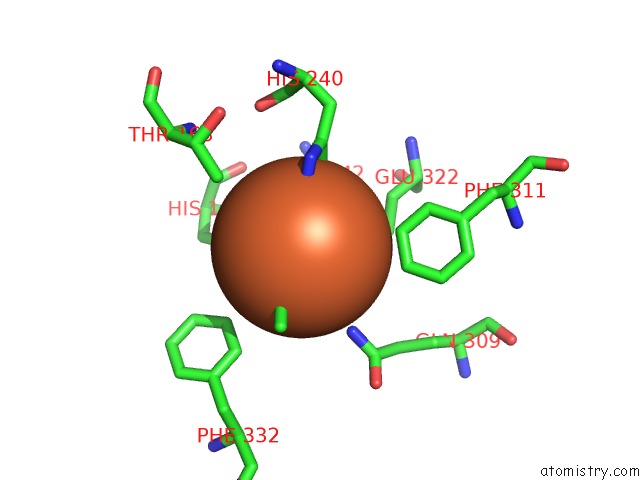
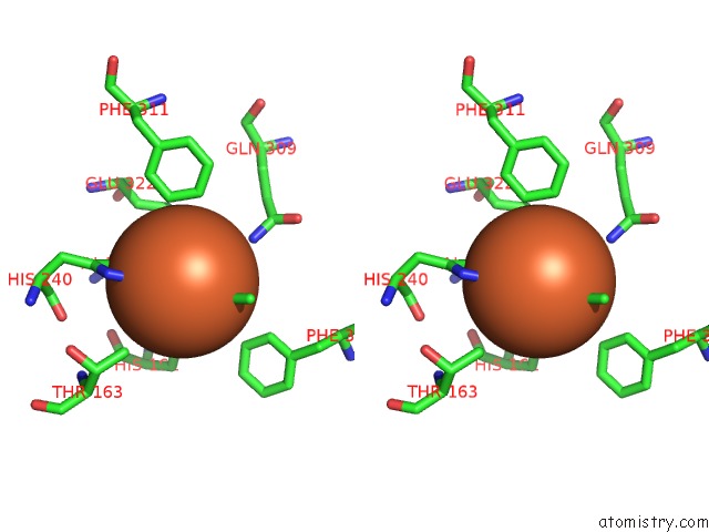
Iron binding site 1 out of 4 in 1cjx
Go back to
Iron binding site 1 out
of 4 in the Crystal Structure of Pseudomonas Fluorescens Hppd
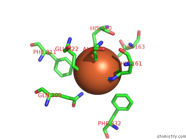
Mono view
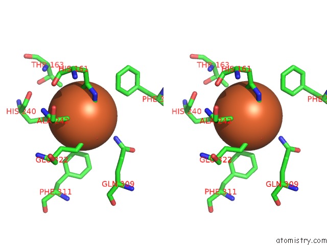
Stereo pair view
Mono view
Stereo pair view
A full contact list of Iron with other atoms in the Fe binding
site number 1 of Crystal Structure of Pseudomonas Fluorescens Hppd within 5.0Å range:
|
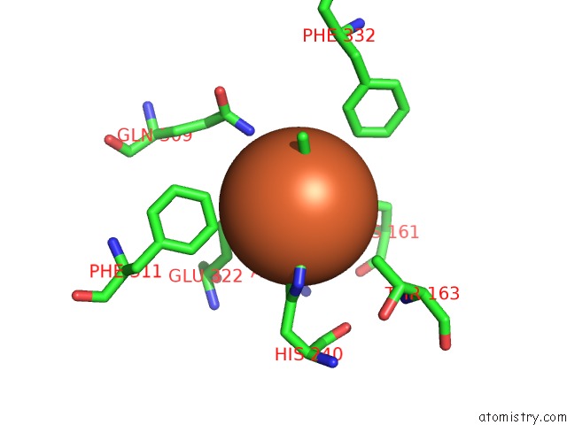
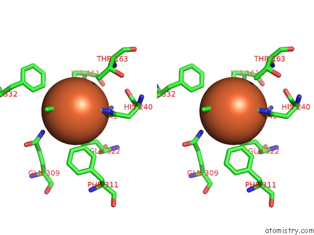
Iron binding site 2 out of 4 in 1cjx
Go back to
Iron binding site 2 out
of 4 in the Crystal Structure of Pseudomonas Fluorescens Hppd
Mono view
Stereo pair view
Mono view
Stereo pair view
A full contact list of Iron with other atoms in the Fe binding
site number 2 of Crystal Structure of Pseudomonas Fluorescens Hppd within 5.0Å range:
|
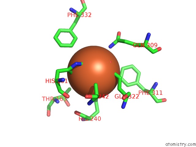
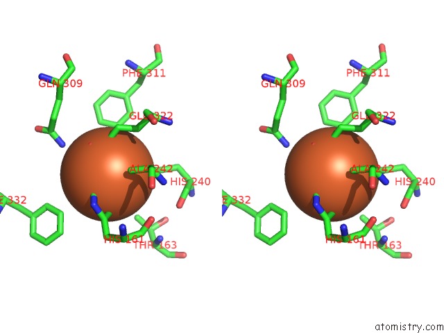
Iron binding site 3 out of 4 in 1cjx
Go back to
Iron binding site 3 out
of 4 in the Crystal Structure of Pseudomonas Fluorescens Hppd
Mono view
Stereo pair view
Mono view
Stereo pair view
A full contact list of Iron with other atoms in the Fe binding
site number 3 of Crystal Structure of Pseudomonas Fluorescens Hppd within 5.0Å range:
|
Iron binding site 4 out of 4 in 1cjx
Go back to
Iron binding site 4 out
of 4 in the Crystal Structure of Pseudomonas Fluorescens Hppd
Mono view
Stereo pair view
Mono view
Stereo pair view
A full contact list of Iron with other atoms in the Fe binding
site number 4 of Crystal Structure of Pseudomonas Fluorescens Hppd within 5.0Å range:
|
Reference:
L.Serre,
A.Sailland,
D.Sy,
P.Boudec,
A.Rolland,
E.Pebay-Peyroula,
C.Cohen-Addad.
Crystal Structure of Pseudomonas Fluorescens 4-Hydroxyphenylpyruvate Dioxygenase: An Enzyme Involved in the Tyrosine Degradation Pathway. Structure Fold.Des. V. 7 977 1999.
ISSN: ISSN 0969-2126
PubMed: 10467142
DOI: 10.1016/S0969-2126(99)80124-5
Page generated: Wed Jul 16 12:59:06 2025
ISSN: ISSN 0969-2126
PubMed: 10467142
DOI: 10.1016/S0969-2126(99)80124-5
Last articles
W in 1DV4W in 1FR3
W in 1GUG
W in 1H9R
W in 1H9K
W in 1H0H
W in 1FEZ
W in 1FKA
W in 1E3P
W in 1E18