Iron »
PDB 1ch3-1cp2 »
1cot »
Iron in PDB 1cot: X-Ray Structure of the Cytochrome C2 Isolated From Paracoccus Denitrificans Refined to 1.7 Angstroms Resolution
Protein crystallography data
The structure of X-Ray Structure of the Cytochrome C2 Isolated From Paracoccus Denitrificans Refined to 1.7 Angstroms Resolution, PDB code: 1cot
was solved by
M.M.Benning,
T.E.Meyer,
H.M.Holden,
with X-Ray Crystallography technique. A brief refinement statistics is given in the table below:
| Resolution Low / High (Å) | 30.00 / 1.70 |
| Space group | P 21 21 21 |
| Cell size a, b, c (Å), α, β, γ (°) | 31.600, 42.100, 81.600, 90.00, 90.00, 90.00 |
| R / Rfree (%) | n/a / n/a |
Iron Binding Sites:
The binding sites of Iron atom in the X-Ray Structure of the Cytochrome C2 Isolated From Paracoccus Denitrificans Refined to 1.7 Angstroms Resolution
(pdb code 1cot). This binding sites where shown within
5.0 Angstroms radius around Iron atom.
In total only one binding site of Iron was determined in the X-Ray Structure of the Cytochrome C2 Isolated From Paracoccus Denitrificans Refined to 1.7 Angstroms Resolution, PDB code: 1cot:
In total only one binding site of Iron was determined in the X-Ray Structure of the Cytochrome C2 Isolated From Paracoccus Denitrificans Refined to 1.7 Angstroms Resolution, PDB code: 1cot:
Iron binding site 1 out of 1 in 1cot
Go back to
Iron binding site 1 out
of 1 in the X-Ray Structure of the Cytochrome C2 Isolated From Paracoccus Denitrificans Refined to 1.7 Angstroms Resolution
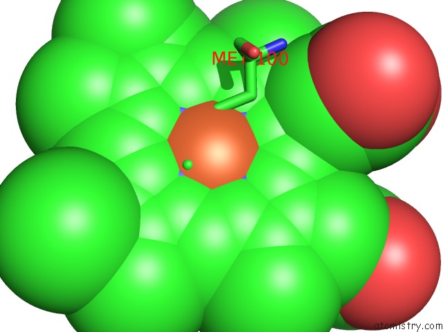
Mono view
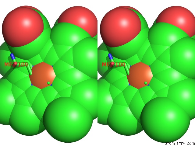
Stereo pair view
Mono view
Stereo pair view
A full contact list of Iron with other atoms in the Fe binding
site number 1 of X-Ray Structure of the Cytochrome C2 Isolated From Paracoccus Denitrificans Refined to 1.7 Angstroms Resolution within 5.0Å range:
|
Reference:
M.M.Benning,
T.E.Meyer,
H.M.Holden.
X-Ray Structure of the Cytochrome C2 Isolated From Paracoccus Denitrificans Refined to 1.7-A Resolution. Arch.Biochem.Biophys. V. 310 460 1994.
ISSN: ISSN 0003-9861
PubMed: 8179333
DOI: 10.1006/ABBI.1994.1193
Page generated: Wed Jul 16 13:03:28 2025
ISSN: ISSN 0003-9861
PubMed: 8179333
DOI: 10.1006/ABBI.1994.1193
Last articles
Fe in 2YXOFe in 2YRS
Fe in 2YXC
Fe in 2YNM
Fe in 2YVJ
Fe in 2YP1
Fe in 2YU2
Fe in 2YU1
Fe in 2YQB
Fe in 2YOO