Iron »
PDB 1cp4-1czp »
1cpq »
Iron in PDB 1cpq: Cytochrome C' From Rhodopseudomonas Capsulata
Protein crystallography data
The structure of Cytochrome C' From Rhodopseudomonas Capsulata, PDB code: 1cpq
was solved by
T.H.Tahirov,
S.Misaki,
T.E.Meyer,
M.A.Cusanovich,
Y.Higuchi,
N.Yasuoka,
with X-Ray Crystallography technique. A brief refinement statistics is given in the table below:
| Resolution Low / High (Å) | 6.00 / 1.72 |
| Space group | P 21 21 2 |
| Cell size a, b, c (Å), α, β, γ (°) | 47.820, 72.590, 34.320, 90.00, 90.00, 90.00 |
| R / Rfree (%) | 15 / n/a |
Iron Binding Sites:
The binding sites of Iron atom in the Cytochrome C' From Rhodopseudomonas Capsulata
(pdb code 1cpq). This binding sites where shown within
5.0 Angstroms radius around Iron atom.
In total only one binding site of Iron was determined in the Cytochrome C' From Rhodopseudomonas Capsulata, PDB code: 1cpq:
In total only one binding site of Iron was determined in the Cytochrome C' From Rhodopseudomonas Capsulata, PDB code: 1cpq:
Iron binding site 1 out of 1 in 1cpq
Go back to
Iron binding site 1 out
of 1 in the Cytochrome C' From Rhodopseudomonas Capsulata
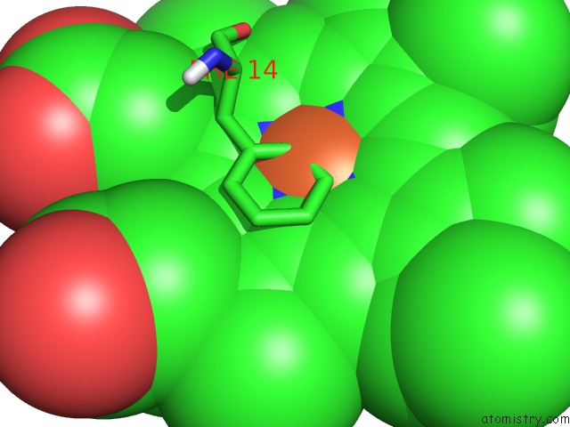
Mono view
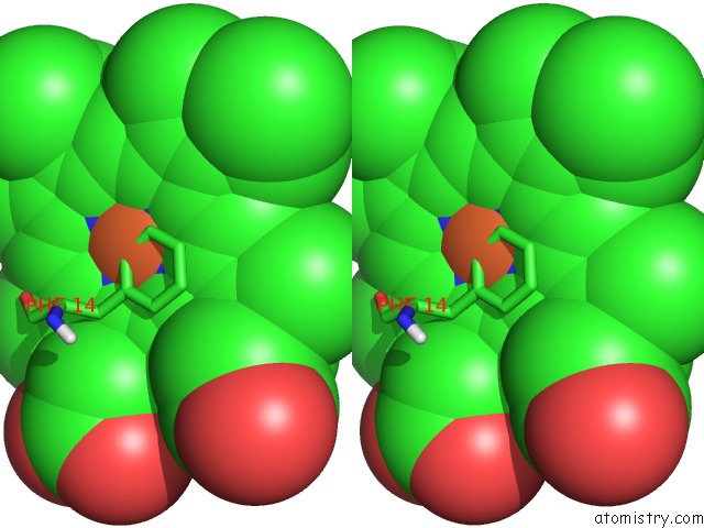
Stereo pair view
Mono view
Stereo pair view
A full contact list of Iron with other atoms in the Fe binding
site number 1 of Cytochrome C' From Rhodopseudomonas Capsulata within 5.0Å range:
|
Reference:
T.H.Tahirov,
S.Misaki,
T.E.Meyer,
M.A.Cusanovich,
Y.Higuchi,
N.Yasuoka.
High-Resolution Crystal Structures of Two Polymorphs of Cytochrome C' From the Purple Phototrophic Bacterium Rhodobacter Capsulatus. J.Mol.Biol. V. 259 467 1996.
ISSN: ISSN 0022-2836
PubMed: 8676382
DOI: 10.1006/JMBI.1996.0333
Page generated: Wed Jul 16 13:04:43 2025
ISSN: ISSN 0022-2836
PubMed: 8676382
DOI: 10.1006/JMBI.1996.0333
Last articles
Zn in 1IRNZn in 1IML
Zn in 1IQB
Zn in 1INN
Zn in 1IQ8
Zn in 1IO0
Zn in 1ILE
Zn in 1IM5
Zn in 1IJL
Zn in 1IBQ