Iron »
PDB 1d06-1dj5 »
1d2v »
Iron in PDB 1d2v: Crystal Structure of Bromide-Bound Human Myeloperoxidase Isoform C at pH 5.5
Enzymatic activity of Crystal Structure of Bromide-Bound Human Myeloperoxidase Isoform C at pH 5.5
All present enzymatic activity of Crystal Structure of Bromide-Bound Human Myeloperoxidase Isoform C at pH 5.5:
1.11.1.7;
1.11.1.7;
Protein crystallography data
The structure of Crystal Structure of Bromide-Bound Human Myeloperoxidase Isoform C at pH 5.5, PDB code: 1d2v
was solved by
T.J.Fiedler,
C.A.Davey,
R.E.Fenna,
with X-Ray Crystallography technique. A brief refinement statistics is given in the table below:
| Resolution Low / High (Å) | 30.00 / 1.75 |
| Space group | P 1 21 1 |
| Cell size a, b, c (Å), α, β, γ (°) | 111.155, 63.488, 92.476, 90.00, 97.36, 90.00 |
| R / Rfree (%) | 24.3 / 29.6 |
Other elements in 1d2v:
The structure of Crystal Structure of Bromide-Bound Human Myeloperoxidase Isoform C at pH 5.5 also contains other interesting chemical elements:
| Bromine | (Br) | 8 atoms |
| Calcium | (Ca) | 2 atoms |
Iron Binding Sites:
The binding sites of Iron atom in the Crystal Structure of Bromide-Bound Human Myeloperoxidase Isoform C at pH 5.5
(pdb code 1d2v). This binding sites where shown within
5.0 Angstroms radius around Iron atom.
In total 2 binding sites of Iron where determined in the Crystal Structure of Bromide-Bound Human Myeloperoxidase Isoform C at pH 5.5, PDB code: 1d2v:
Jump to Iron binding site number: 1; 2;
In total 2 binding sites of Iron where determined in the Crystal Structure of Bromide-Bound Human Myeloperoxidase Isoform C at pH 5.5, PDB code: 1d2v:
Jump to Iron binding site number: 1; 2;
Iron binding site 1 out of 2 in 1d2v
Go back to
Iron binding site 1 out
of 2 in the Crystal Structure of Bromide-Bound Human Myeloperoxidase Isoform C at pH 5.5
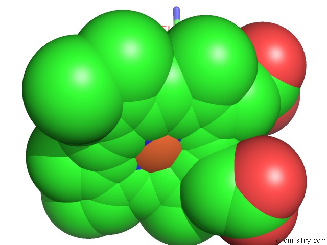
Mono view
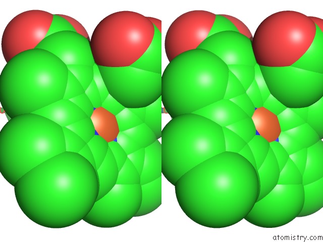
Stereo pair view
Mono view
Stereo pair view
A full contact list of Iron with other atoms in the Fe binding
site number 1 of Crystal Structure of Bromide-Bound Human Myeloperoxidase Isoform C at pH 5.5 within 5.0Å range:
|
Iron binding site 2 out of 2 in 1d2v
Go back to
Iron binding site 2 out
of 2 in the Crystal Structure of Bromide-Bound Human Myeloperoxidase Isoform C at pH 5.5
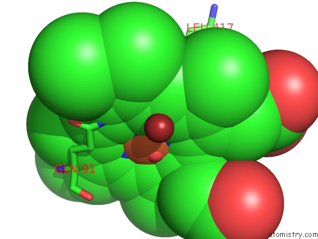
Mono view
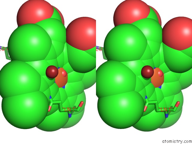
Stereo pair view
Mono view
Stereo pair view
A full contact list of Iron with other atoms in the Fe binding
site number 2 of Crystal Structure of Bromide-Bound Human Myeloperoxidase Isoform C at pH 5.5 within 5.0Å range:
|
Reference:
T.J.Fiedler,
C.A.Davey,
R.E.Fenna.
X-Ray Crystal Structure and Characterization of Halide-Binding Sites of Human Myeloperoxidase at 1.8 A Resolution. J.Biol.Chem. V. 275 11964 2000.
ISSN: ISSN 0021-9258
PubMed: 10766826
DOI: 10.1074/JBC.275.16.11964
Page generated: Wed Jul 16 13:09:51 2025
ISSN: ISSN 0021-9258
PubMed: 10766826
DOI: 10.1074/JBC.275.16.11964
Last articles
K in 5K09K in 5KOE
K in 5KMT
K in 5KIL
K in 5KIK
K in 5KFX
K in 5KGR
K in 5KFW
K in 5KFV
K in 5KFU