Iron »
PDB 1d06-1dj5 »
1d5l »
Iron in PDB 1d5l: Crystal Structure of Cyanide-Bound Human Myeloperoxidase Isoform C at pH 5.5
Enzymatic activity of Crystal Structure of Cyanide-Bound Human Myeloperoxidase Isoform C at pH 5.5
All present enzymatic activity of Crystal Structure of Cyanide-Bound Human Myeloperoxidase Isoform C at pH 5.5:
1.11.1.7;
1.11.1.7;
Protein crystallography data
The structure of Crystal Structure of Cyanide-Bound Human Myeloperoxidase Isoform C at pH 5.5, PDB code: 1d5l
was solved by
T.J.Fiedler,
C.A.Davey,
R.E.Fenna,
with X-Ray Crystallography technique. A brief refinement statistics is given in the table below:
| Resolution Low / High (Å) | 30.00 / 1.90 |
| Space group | P 1 21 1 |
| Cell size a, b, c (Å), α, β, γ (°) | 111.215, 63.507, 92.337, 90.00, 97.43, 90.00 |
| R / Rfree (%) | 17.2 / 21.5 |
Other elements in 1d5l:
The structure of Crystal Structure of Cyanide-Bound Human Myeloperoxidase Isoform C at pH 5.5 also contains other interesting chemical elements:
| Chlorine | (Cl) | 2 atoms |
| Calcium | (Ca) | 2 atoms |
Iron Binding Sites:
The binding sites of Iron atom in the Crystal Structure of Cyanide-Bound Human Myeloperoxidase Isoform C at pH 5.5
(pdb code 1d5l). This binding sites where shown within
5.0 Angstroms radius around Iron atom.
In total 2 binding sites of Iron where determined in the Crystal Structure of Cyanide-Bound Human Myeloperoxidase Isoform C at pH 5.5, PDB code: 1d5l:
Jump to Iron binding site number: 1; 2;
In total 2 binding sites of Iron where determined in the Crystal Structure of Cyanide-Bound Human Myeloperoxidase Isoform C at pH 5.5, PDB code: 1d5l:
Jump to Iron binding site number: 1; 2;
Iron binding site 1 out of 2 in 1d5l
Go back to
Iron binding site 1 out
of 2 in the Crystal Structure of Cyanide-Bound Human Myeloperoxidase Isoform C at pH 5.5
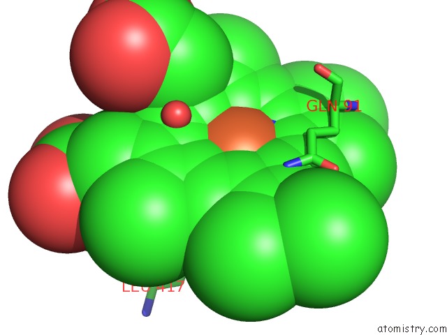
Mono view
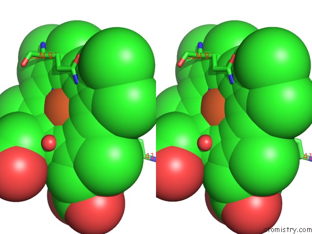
Stereo pair view
Mono view
Stereo pair view
A full contact list of Iron with other atoms in the Fe binding
site number 1 of Crystal Structure of Cyanide-Bound Human Myeloperoxidase Isoform C at pH 5.5 within 5.0Å range:
|
Iron binding site 2 out of 2 in 1d5l
Go back to
Iron binding site 2 out
of 2 in the Crystal Structure of Cyanide-Bound Human Myeloperoxidase Isoform C at pH 5.5
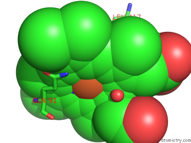
Mono view
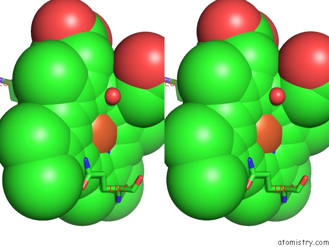
Stereo pair view
Mono view
Stereo pair view
A full contact list of Iron with other atoms in the Fe binding
site number 2 of Crystal Structure of Cyanide-Bound Human Myeloperoxidase Isoform C at pH 5.5 within 5.0Å range:
|
Reference:
M.Blair-Johnson,
T.Fiedler,
R.Fenna.
Human Myeloperoxidase: Structure of A Cyanide Complex and Its Interaction with Bromide and Thiocyanate Substrates at 1.9 A Resolution. Biochemistry V. 40 13990 2001.
ISSN: ISSN 0006-2960
PubMed: 11705390
DOI: 10.1021/BI0111808
Page generated: Wed Jul 16 13:11:45 2025
ISSN: ISSN 0006-2960
PubMed: 11705390
DOI: 10.1021/BI0111808
Last articles
I in 7P4OI in 7PCP
I in 7OIW
I in 7P31
I in 7P2G
I in 7O2R
I in 7OUL
I in 7N4Y
I in 7O2P
I in 7O48