Iron »
PDB 1d06-1dj5 »
1dfx »
Iron in PDB 1dfx: Desulfoferrodoxin From Desulfovibrio Desulfuricans, Atcc 27774
Protein crystallography data
The structure of Desulfoferrodoxin From Desulfovibrio Desulfuricans, Atcc 27774, PDB code: 1dfx
was solved by
A.V.Coelho,
P.M.Matias,
M.A.Carrondo,
with X-Ray Crystallography technique. A brief refinement statistics is given in the table below:
| Resolution Low / High (Å) | 20.00 / 1.90 |
| Space group | H 3 2 |
| Cell size a, b, c (Å), α, β, γ (°) | 112.500, 112.500, 63.200, 90.00, 90.00, 120.00 |
| R / Rfree (%) | 20 / 23 |
Other elements in 1dfx:
The structure of Desulfoferrodoxin From Desulfovibrio Desulfuricans, Atcc 27774 also contains other interesting chemical elements:
| Calcium | (Ca) | 1 atom |
Iron Binding Sites:
The binding sites of Iron atom in the Desulfoferrodoxin From Desulfovibrio Desulfuricans, Atcc 27774
(pdb code 1dfx). This binding sites where shown within
5.0 Angstroms radius around Iron atom.
In total 2 binding sites of Iron where determined in the Desulfoferrodoxin From Desulfovibrio Desulfuricans, Atcc 27774, PDB code: 1dfx:
Jump to Iron binding site number: 1; 2;
In total 2 binding sites of Iron where determined in the Desulfoferrodoxin From Desulfovibrio Desulfuricans, Atcc 27774, PDB code: 1dfx:
Jump to Iron binding site number: 1; 2;
Iron binding site 1 out of 2 in 1dfx
Go back to
Iron binding site 1 out
of 2 in the Desulfoferrodoxin From Desulfovibrio Desulfuricans, Atcc 27774
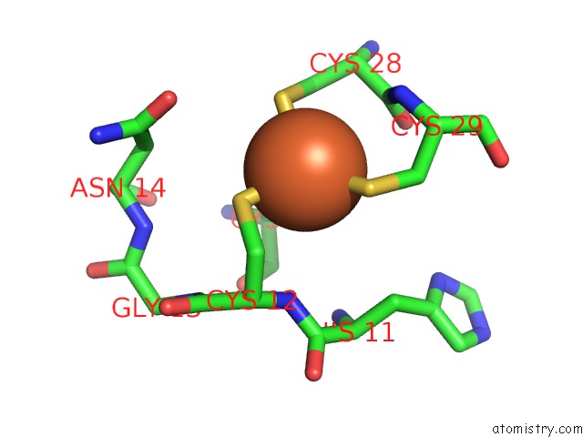
Mono view
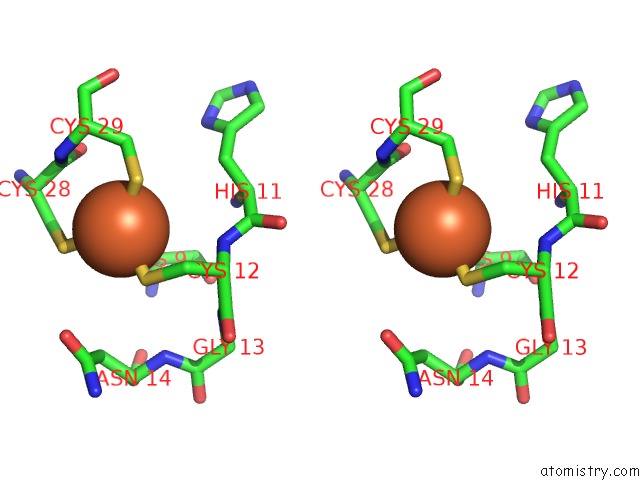
Stereo pair view
Mono view
Stereo pair view
A full contact list of Iron with other atoms in the Fe binding
site number 1 of Desulfoferrodoxin From Desulfovibrio Desulfuricans, Atcc 27774 within 5.0Å range:
|
Iron binding site 2 out of 2 in 1dfx
Go back to
Iron binding site 2 out
of 2 in the Desulfoferrodoxin From Desulfovibrio Desulfuricans, Atcc 27774
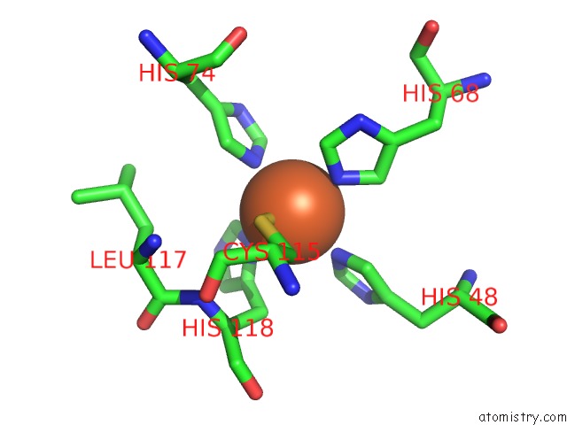
Mono view
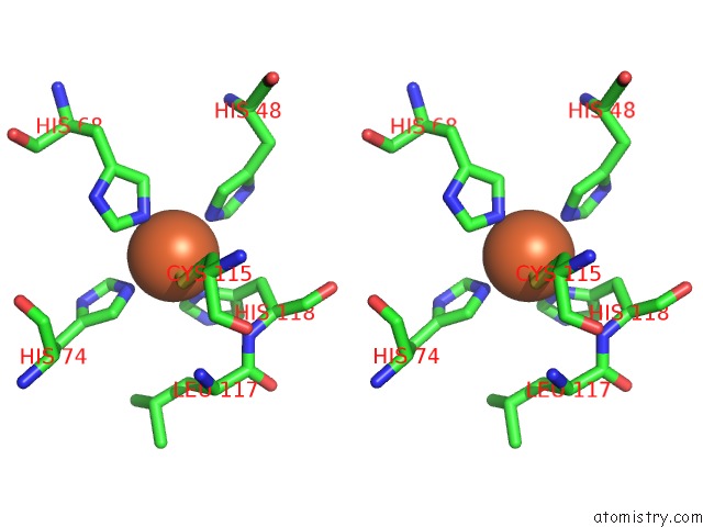
Stereo pair view
Mono view
Stereo pair view
A full contact list of Iron with other atoms in the Fe binding
site number 2 of Desulfoferrodoxin From Desulfovibrio Desulfuricans, Atcc 27774 within 5.0Å range:
|
Reference:
A.V.Coelho,
P.Matias,
V.Fulop,
A.Thompson,
A.Gonzalez,
M.A.Carrondo.
Desulfoferrodoxin Structure Determined By Mad Phasing and Refinement to 1.9 Angstroms Resolution Reveals A Unique Combination of A Tetrahedral FES4 Centre with A Square Pyramidal FESN4 Centre J.Biol.Inorg.Chem. V. 2 680 1997.
ISSN: ISSN 0949-8257
Page generated: Wed Jul 16 13:15:09 2025
ISSN: ISSN 0949-8257
Last articles
K in 7QQRK in 7QQS
K in 7QQQ
K in 7QQP
K in 7QQO
K in 7QK5
K in 7QIX
K in 7QNO
K in 7QIY
K in 7Q3X