Iron »
PDB 1dj7-1dry »
1dkh »
Iron in PDB 1dkh: Crystal Structure of the Hemophore Hasa, pH 6.5
Protein crystallography data
The structure of Crystal Structure of the Hemophore Hasa, pH 6.5, PDB code: 1dkh
was solved by
P.Arnoux,
R.Haser,
N.Izadi-Pruneyre,
A.Lecroisey,
M.Czjzek,
with X-Ray Crystallography technique. A brief refinement statistics is given in the table below:
| Resolution Low / High (Å) | 12.00 / 3.20 |
| Space group | P 32 2 1 |
| Cell size a, b, c (Å), α, β, γ (°) | 111.650, 111.650, 52.790, 90.00, 90.00, 120.00 |
| R / Rfree (%) | 22.8 / 28.8 |
Other elements in 1dkh:
The structure of Crystal Structure of the Hemophore Hasa, pH 6.5 also contains other interesting chemical elements:
| Samarium | (Sm) | 1 atom |
| Zinc | (Zn) | 3 atoms |
Iron Binding Sites:
The binding sites of Iron atom in the Crystal Structure of the Hemophore Hasa, pH 6.5
(pdb code 1dkh). This binding sites where shown within
5.0 Angstroms radius around Iron atom.
In total only one binding site of Iron was determined in the Crystal Structure of the Hemophore Hasa, pH 6.5, PDB code: 1dkh:
In total only one binding site of Iron was determined in the Crystal Structure of the Hemophore Hasa, pH 6.5, PDB code: 1dkh:
Iron binding site 1 out of 1 in 1dkh
Go back to
Iron binding site 1 out
of 1 in the Crystal Structure of the Hemophore Hasa, pH 6.5
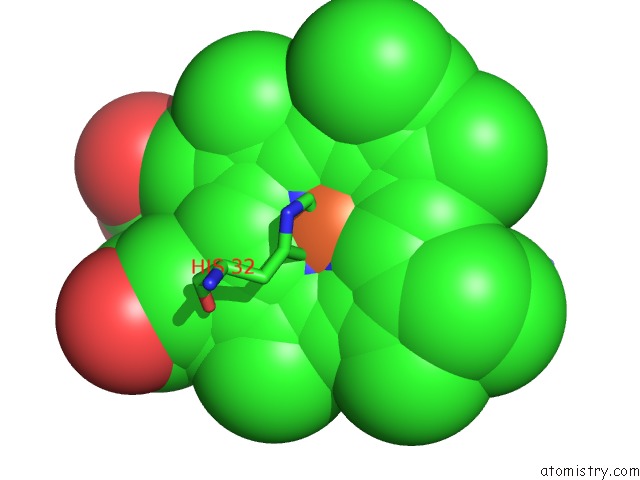
Mono view
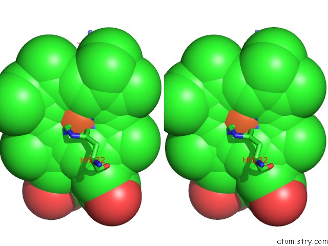
Stereo pair view
Mono view
Stereo pair view
A full contact list of Iron with other atoms in the Fe binding
site number 1 of Crystal Structure of the Hemophore Hasa, pH 6.5 within 5.0Å range:
|
Reference:
P.Arnoux,
R.Haser,
N.Izadi-Pruneyre,
A.Lecroisey,
M.Czjzek.
Functional Aspects of the Heme Bound Hemophore Hasa By Structural Analysis of Various Crystal Forms. Proteins V. 41 202 2000.
ISSN: ISSN 0887-3585
PubMed: 10966573
DOI: 10.1002/1097-0134(20001101)41:2<202::AID-PROT50>3.0.CO;2-8
Page generated: Wed Jul 16 13:19:08 2025
ISSN: ISSN 0887-3585
PubMed: 10966573
DOI: 10.1002/1097-0134(20001101)41:2<202::AID-PROT50>3.0.CO;2-8
Last articles
Mn in 9LJUMn in 9LJW
Mn in 9LJS
Mn in 9LJR
Mn in 9LJT
Mn in 9LJV
Mg in 9UA2
Mg in 9R96
Mg in 9VM1
Mg in 9P01