Iron »
PDB 1dj7-1dry »
1dm1 »
Iron in PDB 1dm1: 2.0 A Crystal Structure of the Double Mutant H(E7)V, T(E10) R of Myoglobin From Aplysia Limacina
Protein crystallography data
The structure of 2.0 A Crystal Structure of the Double Mutant H(E7)V, T(E10) R of Myoglobin From Aplysia Limacina, PDB code: 1dm1
was solved by
L.Federici,
C.Savino,
R.Musto,
C.Travaglini-Allocatelli,
F.Cutruzzola,
M.Brunori,
with X-Ray Crystallography technique. A brief refinement statistics is given in the table below:
| Resolution Low / High (Å) | 59.76 / 1.99 |
| Space group | H 3 |
| Cell size a, b, c (Å), α, β, γ (°) | 89.730, 89.730, 92.080, 90.00, 90.00, 120.00 |
| R / Rfree (%) | 18.9 / 21.6 |
Iron Binding Sites:
The binding sites of Iron atom in the 2.0 A Crystal Structure of the Double Mutant H(E7)V, T(E10) R of Myoglobin From Aplysia Limacina
(pdb code 1dm1). This binding sites where shown within
5.0 Angstroms radius around Iron atom.
In total only one binding site of Iron was determined in the 2.0 A Crystal Structure of the Double Mutant H(E7)V, T(E10) R of Myoglobin From Aplysia Limacina, PDB code: 1dm1:
In total only one binding site of Iron was determined in the 2.0 A Crystal Structure of the Double Mutant H(E7)V, T(E10) R of Myoglobin From Aplysia Limacina, PDB code: 1dm1:
Iron binding site 1 out of 1 in 1dm1
Go back to
Iron binding site 1 out
of 1 in the 2.0 A Crystal Structure of the Double Mutant H(E7)V, T(E10) R of Myoglobin From Aplysia Limacina
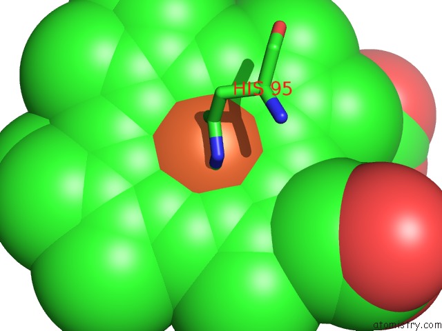
Mono view
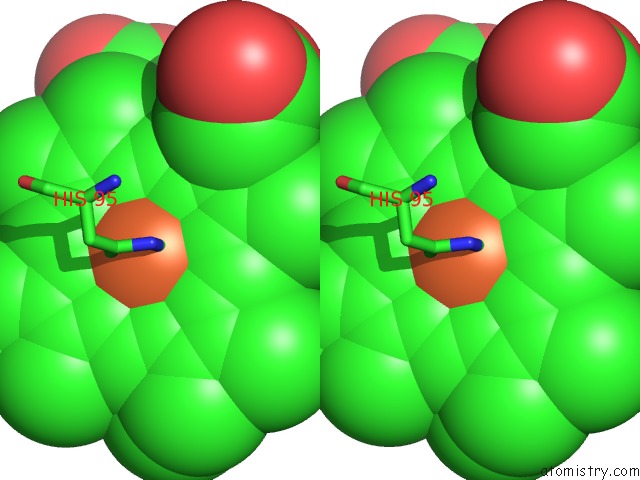
Stereo pair view
Mono view
Stereo pair view
A full contact list of Iron with other atoms in the Fe binding
site number 1 of 2.0 A Crystal Structure of the Double Mutant H(E7)V, T(E10) R of Myoglobin From Aplysia Limacina within 5.0Å range:
|
Reference:
L.Federici,
C.Savino,
R.Musto,
C.Travaglini-Allocatelli,
F.Cutruzzola,
M.Brunori.
Engineering His(E7) Affects the Control of Heme Reactivity in Aplysia Limacina Myoglobin. Biochem.Biophys.Res.Commun. V. 269 58 2000.
ISSN: ISSN 0006-291X
PubMed: 10694477
DOI: 10.1006/BBRC.2000.2259
Page generated: Wed Jul 16 13:20:23 2025
ISSN: ISSN 0006-291X
PubMed: 10694477
DOI: 10.1006/BBRC.2000.2259
Last articles
I in 9ET8I in 9ET1
I in 9ESZ
I in 9E56
I in 9DXP
I in 9E4X
I in 9E50
I in 9DQ1
I in 9DWY
I in 9DAJ