Iron »
PDB 1dj7-1dry »
1do7 »
Iron in PDB 1do7: Carbonmonoxy-Myoglobin (Mutant L29W) Rebinding Structure After Photolysis at T< 180K
Protein crystallography data
The structure of Carbonmonoxy-Myoglobin (Mutant L29W) Rebinding Structure After Photolysis at T< 180K, PDB code: 1do7
was solved by
A.Ostermann,
R.Waschipky,
F.G.Parak,
G.U.Nienhaus,
with X-Ray Crystallography technique. A brief refinement statistics is given in the table below:
| Resolution Low / High (Å) | 7.00 / 1.85 |
| Space group | P 6 |
| Cell size a, b, c (Å), α, β, γ (°) | 90.430, 90.430, 45.240, 90.00, 90.00, 120.00 |
| R / Rfree (%) | 18.6 / 21.4 |
Iron Binding Sites:
The binding sites of Iron atom in the Carbonmonoxy-Myoglobin (Mutant L29W) Rebinding Structure After Photolysis at T< 180K
(pdb code 1do7). This binding sites where shown within
5.0 Angstroms radius around Iron atom.
In total only one binding site of Iron was determined in the Carbonmonoxy-Myoglobin (Mutant L29W) Rebinding Structure After Photolysis at T< 180K, PDB code: 1do7:
In total only one binding site of Iron was determined in the Carbonmonoxy-Myoglobin (Mutant L29W) Rebinding Structure After Photolysis at T< 180K, PDB code: 1do7:
Iron binding site 1 out of 1 in 1do7
Go back to
Iron binding site 1 out
of 1 in the Carbonmonoxy-Myoglobin (Mutant L29W) Rebinding Structure After Photolysis at T< 180K
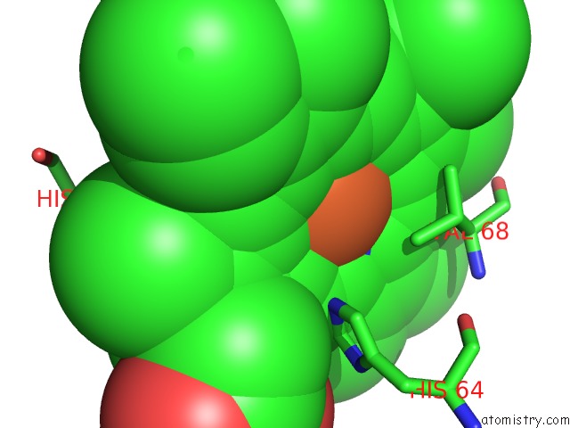
Mono view
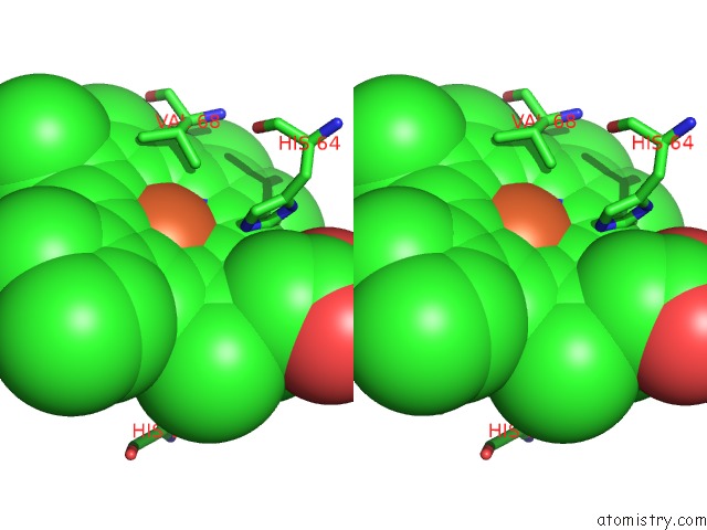
Stereo pair view
Mono view
Stereo pair view
A full contact list of Iron with other atoms in the Fe binding
site number 1 of Carbonmonoxy-Myoglobin (Mutant L29W) Rebinding Structure After Photolysis at T< 180K within 5.0Å range:
|
Reference:
A.Ostermann,
R.Waschipky,
F.G.Parak,
G.U.Nienhaus.
Ligand Binding and Conformational Motions in Myoglobin. Nature V. 404 205 2000.
ISSN: ISSN 0028-0836
PubMed: 10724176
DOI: 10.1038/35004622
Page generated: Wed Jul 16 13:22:55 2025
ISSN: ISSN 0028-0836
PubMed: 10724176
DOI: 10.1038/35004622
Last articles
I in 8U9FI in 8XB3
I in 8X17
I in 8X16
I in 8WGU
I in 8WGT
I in 8W5B
I in 8W17
I in 8VHD
I in 8W16