Iron »
PDB 1ds1-1dxr »
1dsg »
Iron in PDB 1dsg: Cytochrome C Peroxidase H175G Mutant, Imidazole Complex at pH 5, Room Temperature.
Enzymatic activity of Cytochrome C Peroxidase H175G Mutant, Imidazole Complex at pH 5, Room Temperature.
All present enzymatic activity of Cytochrome C Peroxidase H175G Mutant, Imidazole Complex at pH 5, Room Temperature.:
1.11.1.5;
1.11.1.5;
Protein crystallography data
The structure of Cytochrome C Peroxidase H175G Mutant, Imidazole Complex at pH 5, Room Temperature., PDB code: 1dsg
was solved by
J.Hirst,
S.K.Wilcox,
P.A.Williams,
D.E.Mcree,
D.B.Goodin,
with X-Ray Crystallography technique. A brief refinement statistics is given in the table below:
| Resolution Low / High (Å) | 11.00 / 2.56 |
| Space group | P 21 21 21 |
| Cell size a, b, c (Å), α, β, γ (°) | 107.900, 76.610, 51.470, 90.00, 90.00, 90.00 |
| R / Rfree (%) | 15.7 / n/a |
Iron Binding Sites:
The binding sites of Iron atom in the Cytochrome C Peroxidase H175G Mutant, Imidazole Complex at pH 5, Room Temperature.
(pdb code 1dsg). This binding sites where shown within
5.0 Angstroms radius around Iron atom.
In total only one binding site of Iron was determined in the Cytochrome C Peroxidase H175G Mutant, Imidazole Complex at pH 5, Room Temperature., PDB code: 1dsg:
In total only one binding site of Iron was determined in the Cytochrome C Peroxidase H175G Mutant, Imidazole Complex at pH 5, Room Temperature., PDB code: 1dsg:
Iron binding site 1 out of 1 in 1dsg
Go back to
Iron binding site 1 out
of 1 in the Cytochrome C Peroxidase H175G Mutant, Imidazole Complex at pH 5, Room Temperature.
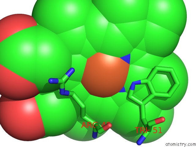
Mono view
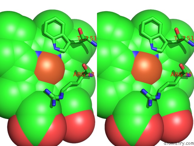
Stereo pair view
Mono view
Stereo pair view
A full contact list of Iron with other atoms in the Fe binding
site number 1 of Cytochrome C Peroxidase H175G Mutant, Imidazole Complex at pH 5, Room Temperature. within 5.0Å range:
|
Reference:
J.Hirst,
S.K.Wilcox,
P.A.Williams,
J.Blankenship,
D.E.Mcree,
D.B.Goodin.
Replacement of the Axial Histidine Ligand with Imidazole in Cytochrome C Peroxidase. 1. Effects on Structure. Biochemistry V. 40 1265 2001.
ISSN: ISSN 0006-2960
PubMed: 11170452
DOI: 10.1021/BI002089R
Page generated: Wed Jul 16 13:25:05 2025
ISSN: ISSN 0006-2960
PubMed: 11170452
DOI: 10.1021/BI002089R
Last articles
Fe in 2YXOFe in 2YRS
Fe in 2YXC
Fe in 2YNM
Fe in 2YVJ
Fe in 2YP1
Fe in 2YU2
Fe in 2YU1
Fe in 2YQB
Fe in 2YOO