Iron »
PDB 1ds1-1dxr »
1dve »
Iron in PDB 1dve: Crystal Structure of Rat Heme Oxygenase-1 in Complex with Heme
Enzymatic activity of Crystal Structure of Rat Heme Oxygenase-1 in Complex with Heme
All present enzymatic activity of Crystal Structure of Rat Heme Oxygenase-1 in Complex with Heme:
1.14.99.3;
1.14.99.3;
Protein crystallography data
The structure of Crystal Structure of Rat Heme Oxygenase-1 in Complex with Heme, PDB code: 1dve
was solved by
M.Sugishima,
Y.Omata,
Y.Kakuta,
H.Sakamoto,
M.Noguchi,
K.Fukuyama,
with X-Ray Crystallography technique. A brief refinement statistics is given in the table below:
| Resolution Low / High (Å) | 27.75 / 2.40 |
| Space group | P 43 21 2 |
| Cell size a, b, c (Å), α, β, γ (°) | 56.139, 56.139, 183.984, 90.00, 90.00, 90.00 |
| R / Rfree (%) | 21.4 / 26.8 |
Iron Binding Sites:
The binding sites of Iron atom in the Crystal Structure of Rat Heme Oxygenase-1 in Complex with Heme
(pdb code 1dve). This binding sites where shown within
5.0 Angstroms radius around Iron atom.
In total only one binding site of Iron was determined in the Crystal Structure of Rat Heme Oxygenase-1 in Complex with Heme, PDB code: 1dve:
In total only one binding site of Iron was determined in the Crystal Structure of Rat Heme Oxygenase-1 in Complex with Heme, PDB code: 1dve:
Iron binding site 1 out of 1 in 1dve
Go back to
Iron binding site 1 out
of 1 in the Crystal Structure of Rat Heme Oxygenase-1 in Complex with Heme
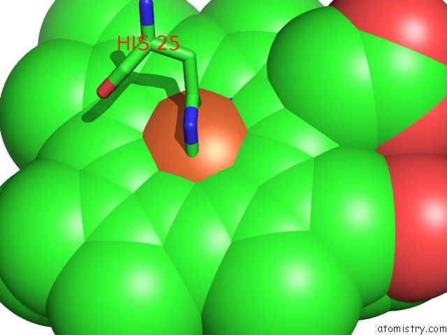
Mono view
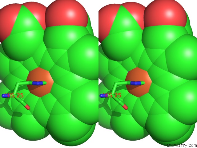
Stereo pair view
Mono view
Stereo pair view
A full contact list of Iron with other atoms in the Fe binding
site number 1 of Crystal Structure of Rat Heme Oxygenase-1 in Complex with Heme within 5.0Å range:
|
Reference:
M.Sugishima,
Y.Omata,
Y.Kakuta,
H.Sakamoto,
M.Noguchi,
K.Fukuyama.
Crystal Structure of Rat Heme Oxygenase-1 in Complex with Heme. Febs Lett. V. 471 61 2000.
ISSN: ISSN 0014-5793
PubMed: 10760513
DOI: 10.1016/S0014-5793(00)01353-3
Page generated: Wed Jul 16 13:26:55 2025
ISSN: ISSN 0014-5793
PubMed: 10760513
DOI: 10.1016/S0014-5793(00)01353-3
Last articles
Na in 3BH4Na in 3BHD
Na in 3BH9
Na in 3B8X
Na in 3BEU
Na in 3BFT
Na in 3BE8
Na in 3BCD
Na in 3BCF
Na in 3BAX