Iron »
PDB 1ds1-1dxr »
1dwt »
Iron in PDB 1dwt: Photorelaxed Horse Heart Myoglobin Co Complex
Protein crystallography data
The structure of Photorelaxed Horse Heart Myoglobin Co Complex, PDB code: 1dwt
was solved by
K.Chu,
J.Vojtechovsky,
B.H.Mcmahon,
R.M.Sweet,
J.Berendzen,
I.Schlichting,
with X-Ray Crystallography technique. A brief refinement statistics is given in the table below:
| Resolution Low / High (Å) | 20.00 / 1.40 |
| Space group | P 1 21 1 |
| Cell size a, b, c (Å), α, β, γ (°) | 62.800, 28.800, 35.200, 90.00, 106.20, 90.00 |
| R / Rfree (%) | 19.7 / 24.3 |
Iron Binding Sites:
The binding sites of Iron atom in the Photorelaxed Horse Heart Myoglobin Co Complex
(pdb code 1dwt). This binding sites where shown within
5.0 Angstroms radius around Iron atom.
In total only one binding site of Iron was determined in the Photorelaxed Horse Heart Myoglobin Co Complex, PDB code: 1dwt:
In total only one binding site of Iron was determined in the Photorelaxed Horse Heart Myoglobin Co Complex, PDB code: 1dwt:
Iron binding site 1 out of 1 in 1dwt
Go back to
Iron binding site 1 out
of 1 in the Photorelaxed Horse Heart Myoglobin Co Complex
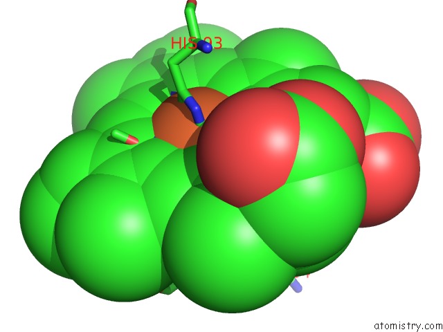
Mono view
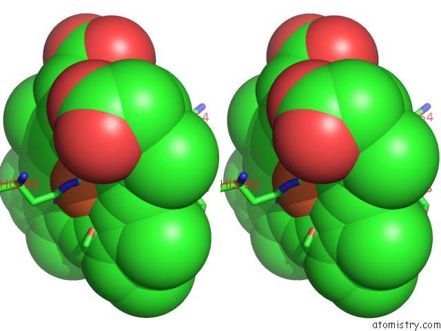
Stereo pair view
Mono view
Stereo pair view
A full contact list of Iron with other atoms in the Fe binding
site number 1 of Photorelaxed Horse Heart Myoglobin Co Complex within 5.0Å range:
|
Reference:
K.Chu,
J.Vojtechovsky,
B.H.Mcmahon,
R.M.Sweet,
J.Berendzen,
I.Schlichting.
Crystal Structure of A New Ligand Binding Intermediate in Wildtype Carbonmonoxy Myoglobin Nature V. 403 921 2000.
ISSN: ISSN 0028-0836
PubMed: 10706294
DOI: 10.1038/35002641
Page generated: Wed Jul 16 13:29:47 2025
ISSN: ISSN 0028-0836
PubMed: 10706294
DOI: 10.1038/35002641
Last articles
Fe in 2YXOFe in 2YRS
Fe in 2YXC
Fe in 2YNM
Fe in 2YVJ
Fe in 2YP1
Fe in 2YU2
Fe in 2YU1
Fe in 2YQB
Fe in 2YOO