Iron »
PDB 1ds1-1dxr »
1dxd »
Iron in PDB 1dxd: Photolyzed Co Complex of Myoglobin Mb-Yqr at 20K
Protein crystallography data
The structure of Photolyzed Co Complex of Myoglobin Mb-Yqr at 20K, PDB code: 1dxd
was solved by
M.Brunori,
B.Vallone,
F.Cutruzzola,
C.Travaglini-Allocatelli,
J.Berendzen,
K.Chu,
R.M.Sweet,
I.Schlichting,
with X-Ray Crystallography technique. A brief refinement statistics is given in the table below:
| Resolution Low / High (Å) | 18 / 1.4 |
| Space group | P 6 |
| Cell size a, b, c (Å), α, β, γ (°) | 90.493, 90.493, 45.283, 90.00, 90.00, 120.00 |
| R / Rfree (%) | 13.3 / 16.9 |
Iron Binding Sites:
The binding sites of Iron atom in the Photolyzed Co Complex of Myoglobin Mb-Yqr at 20K
(pdb code 1dxd). This binding sites where shown within
5.0 Angstroms radius around Iron atom.
In total only one binding site of Iron was determined in the Photolyzed Co Complex of Myoglobin Mb-Yqr at 20K, PDB code: 1dxd:
In total only one binding site of Iron was determined in the Photolyzed Co Complex of Myoglobin Mb-Yqr at 20K, PDB code: 1dxd:
Iron binding site 1 out of 1 in 1dxd
Go back to
Iron binding site 1 out
of 1 in the Photolyzed Co Complex of Myoglobin Mb-Yqr at 20K
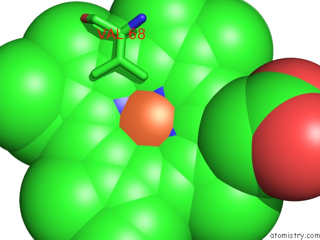
Mono view
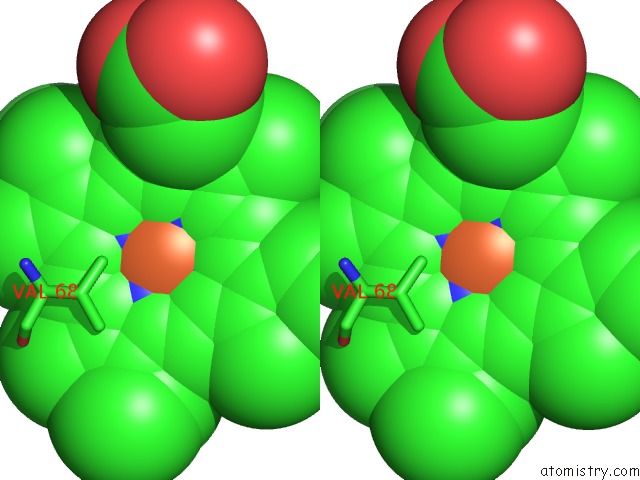
Stereo pair view
Mono view
Stereo pair view
A full contact list of Iron with other atoms in the Fe binding
site number 1 of Photolyzed Co Complex of Myoglobin Mb-Yqr at 20K within 5.0Å range:
|
Reference:
M.Brunori,
B.Vallone,
F.Cutruzzola,
C.Travaglini-Allocatelli,
J.Berendzen,
K.Chu,
R.M.Sweet,
I.Schlichting.
The Role of Cavities in Protein Dynamics: Crystal Structure of A Photolytic Intermediate of A Mutant Myoglobin. Proc.Natl.Acad.Sci.Usa V. 97 2058 2000.
ISSN: ISSN 0027-8424
PubMed: 10681426
DOI: 10.1073/PNAS.040459697
Page generated: Wed Jul 16 13:30:41 2025
ISSN: ISSN 0027-8424
PubMed: 10681426
DOI: 10.1073/PNAS.040459697
Last articles
Na in 3BH9Na in 3B8X
Na in 3BEU
Na in 3BFT
Na in 3BE8
Na in 3BCD
Na in 3BCF
Na in 3BAX
Na in 3BAW
Na in 3BC9