Iron »
PDB 1dxt-1ea1 »
1dyt »
Iron in PDB 1dyt: X-Ray Crystal Structure of Ecp (Rnase 3) at 1.75 A
Protein crystallography data
The structure of X-Ray Crystal Structure of Ecp (Rnase 3) at 1.75 A, PDB code: 1dyt
was solved by
G.Mallorqui-Fernandez,
J.Pous,
R.Peracaula,
T.Maeda,
H.Tada,
H.Yamada,
M.Seno,
R.De Llorens,
F.X.Gomis-Rueth,
M.Coll,
with X-Ray Crystallography technique. A brief refinement statistics is given in the table below:
| Resolution Low / High (Å) | 20 / 1.75 |
| Space group | P 43 2 2 |
| Cell size a, b, c (Å), α, β, γ (°) | 62.163, 62.163, 174.590, 90.00, 90.00, 90.00 |
| R / Rfree (%) | 22.4 / 27.1 |
Iron Binding Sites:
The binding sites of Iron atom in the X-Ray Crystal Structure of Ecp (Rnase 3) at 1.75 A
(pdb code 1dyt). This binding sites where shown within
5.0 Angstroms radius around Iron atom.
In total 2 binding sites of Iron where determined in the X-Ray Crystal Structure of Ecp (Rnase 3) at 1.75 A, PDB code: 1dyt:
Jump to Iron binding site number: 1; 2;
In total 2 binding sites of Iron where determined in the X-Ray Crystal Structure of Ecp (Rnase 3) at 1.75 A, PDB code: 1dyt:
Jump to Iron binding site number: 1; 2;
Iron binding site 1 out of 2 in 1dyt
Go back to
Iron binding site 1 out
of 2 in the X-Ray Crystal Structure of Ecp (Rnase 3) at 1.75 A
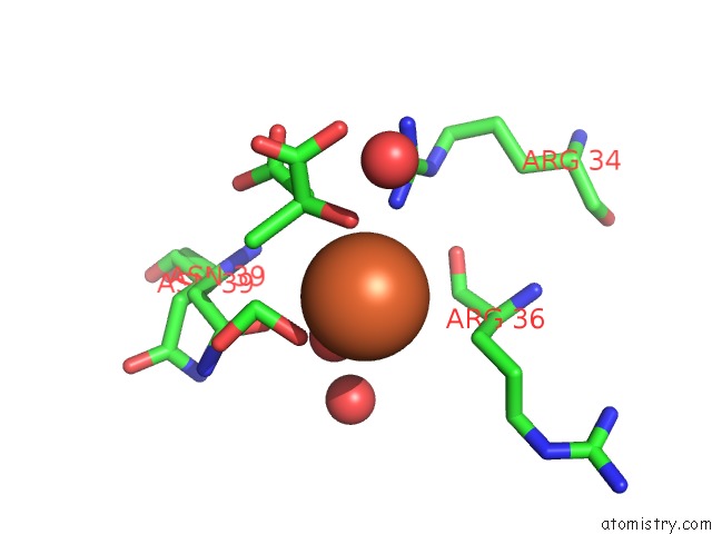
Mono view
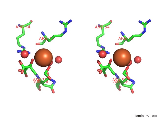
Stereo pair view
Mono view
Stereo pair view
A full contact list of Iron with other atoms in the Fe binding
site number 1 of X-Ray Crystal Structure of Ecp (Rnase 3) at 1.75 A within 5.0Å range:
|
Iron binding site 2 out of 2 in 1dyt
Go back to
Iron binding site 2 out
of 2 in the X-Ray Crystal Structure of Ecp (Rnase 3) at 1.75 A
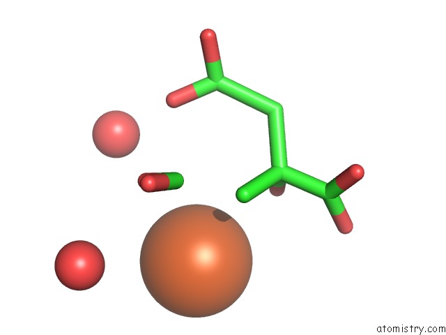
Mono view
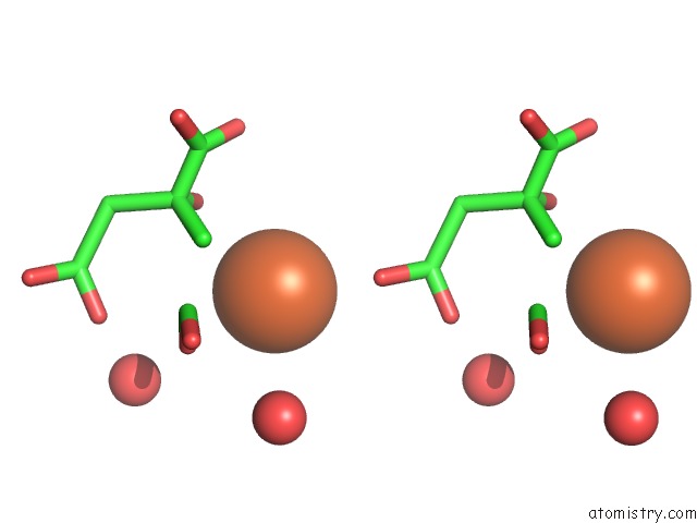
Stereo pair view
Mono view
Stereo pair view
A full contact list of Iron with other atoms in the Fe binding
site number 2 of X-Ray Crystal Structure of Ecp (Rnase 3) at 1.75 A within 5.0Å range:
|
Reference:
G.Mallorqui-Fernandez,
J.Pous,
R.Peracaula,
T.Maeda,
H.Tada,
H.Yamada,
M.Seno,
R.De Llorens,
F.X.Gomis-Rueth,
M.Coll.
Three-Dimensional Crystal Structure of Human Eosinophil Cationic Protein (Rnase 3) at 1.75 A Resolution. J.Mol.Biol. V. 300 1297 2000.
ISSN: ISSN 0022-2836
PubMed: 10903870
DOI: 10.1006/JMBI.2000.3939
Page generated: Wed Jul 16 13:32:23 2025
ISSN: ISSN 0022-2836
PubMed: 10903870
DOI: 10.1006/JMBI.2000.3939
Last articles
K in 4R33K in 4R2C
K in 4QXG
K in 4QRH
K in 4QNE
K in 4QGC
K in 4QKA
K in 4QE9
K in 4QG8
K in 4QK8