Iron »
PDB 1eb7-1esz »
1eb7 »
Iron in PDB 1eb7: Crystal Structure of the Di-Haem Cytochrome C Peroxidase From Pseudomonas Aeruginosa
Enzymatic activity of Crystal Structure of the Di-Haem Cytochrome C Peroxidase From Pseudomonas Aeruginosa
All present enzymatic activity of Crystal Structure of the Di-Haem Cytochrome C Peroxidase From Pseudomonas Aeruginosa:
1.11.1.5;
1.11.1.5;
Protein crystallography data
The structure of Crystal Structure of the Di-Haem Cytochrome C Peroxidase From Pseudomonas Aeruginosa, PDB code: 1eb7
was solved by
V.Fulop,
with X-Ray Crystallography technique. A brief refinement statistics is given in the table below:
| Resolution Low / High (Å) | 25.0 / 2.4 |
| Space group | P 31 2 1 |
| Cell size a, b, c (Å), α, β, γ (°) | 113.900, 113.900, 72.000, 90.00, 90.00, 120.00 |
| R / Rfree (%) | 17.2 / 22 |
Other elements in 1eb7:
The structure of Crystal Structure of the Di-Haem Cytochrome C Peroxidase From Pseudomonas Aeruginosa also contains other interesting chemical elements:
| Calcium | (Ca) | 1 atom |
Iron Binding Sites:
The binding sites of Iron atom in the Crystal Structure of the Di-Haem Cytochrome C Peroxidase From Pseudomonas Aeruginosa
(pdb code 1eb7). This binding sites where shown within
5.0 Angstroms radius around Iron atom.
In total 2 binding sites of Iron where determined in the Crystal Structure of the Di-Haem Cytochrome C Peroxidase From Pseudomonas Aeruginosa, PDB code: 1eb7:
Jump to Iron binding site number: 1; 2;
In total 2 binding sites of Iron where determined in the Crystal Structure of the Di-Haem Cytochrome C Peroxidase From Pseudomonas Aeruginosa, PDB code: 1eb7:
Jump to Iron binding site number: 1; 2;
Iron binding site 1 out of 2 in 1eb7
Go back to
Iron binding site 1 out
of 2 in the Crystal Structure of the Di-Haem Cytochrome C Peroxidase From Pseudomonas Aeruginosa
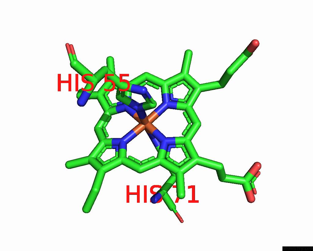
Mono view
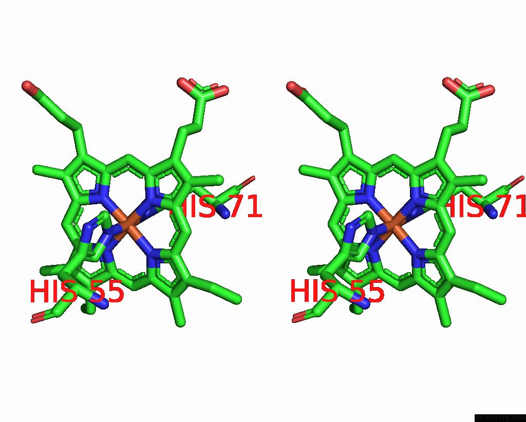
Stereo pair view
Mono view
Stereo pair view
A full contact list of Iron with other atoms in the Fe binding
site number 1 of Crystal Structure of the Di-Haem Cytochrome C Peroxidase From Pseudomonas Aeruginosa within 5.0Å range:
|
Iron binding site 2 out of 2 in 1eb7
Go back to
Iron binding site 2 out
of 2 in the Crystal Structure of the Di-Haem Cytochrome C Peroxidase From Pseudomonas Aeruginosa
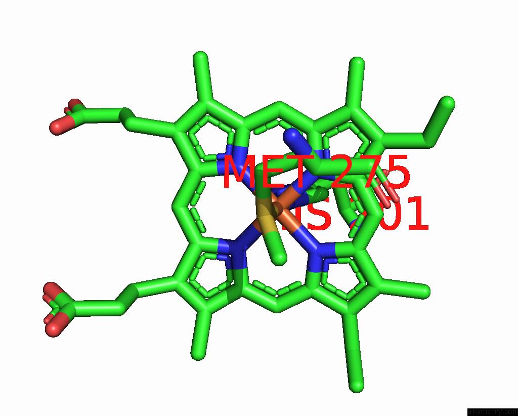
Mono view
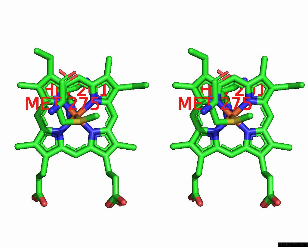
Stereo pair view
Mono view
Stereo pair view
A full contact list of Iron with other atoms in the Fe binding
site number 2 of Crystal Structure of the Di-Haem Cytochrome C Peroxidase From Pseudomonas Aeruginosa within 5.0Å range:
|
Reference:
V.Fulop,
C.J.Ridout,
C.Greenwood,
J.Hajdu.
Crystal Structure of the Di-Haemcytochrome C Peroxidase From Pseudomonas Aeruginosa Structure V. 3 1225 1995.
ISSN: ISSN 0969-2126
PubMed: 8591033
DOI: 10.1016/S0969-2126(01)00258-1
Page generated: Wed Jul 16 13:47:15 2025
ISSN: ISSN 0969-2126
PubMed: 8591033
DOI: 10.1016/S0969-2126(01)00258-1
Last articles
I in 6AXTI in 6AXV
I in 6AXS
I in 6AN0
I in 6AXR
I in 5ZBA
I in 6A4Y
I in 5ZRF
I in 6A9B
I in 6A9A