Iron »
PDB 1eb7-1esz »
1ehb »
Iron in PDB 1ehb: Crystal Structure of Recombinant Trypsin-Solubilized Fragment of Cytochrome B5
Protein crystallography data
The structure of Crystal Structure of Recombinant Trypsin-Solubilized Fragment of Cytochrome B5, PDB code: 1ehb
was solved by
J.Wu,
J.-H.Gan,
Z.-X.Xia,
Y.-H.Wang,
W.-H.Wang,
L.-L.Xue,
Y.Xie,
Z.-X.Huang,
with X-Ray Crystallography technique. A brief refinement statistics is given in the table below:
| Resolution Low / High (Å) | 100.00 / 1.90 |
| Space group | C 1 2 1 |
| Cell size a, b, c (Å), α, β, γ (°) | 70.700, 40.440, 39.280, 90.00, 111.76, 90.00 |
| R / Rfree (%) | 19.8 / 24.8 |
Iron Binding Sites:
The binding sites of Iron atom in the Crystal Structure of Recombinant Trypsin-Solubilized Fragment of Cytochrome B5
(pdb code 1ehb). This binding sites where shown within
5.0 Angstroms radius around Iron atom.
In total only one binding site of Iron was determined in the Crystal Structure of Recombinant Trypsin-Solubilized Fragment of Cytochrome B5, PDB code: 1ehb:
In total only one binding site of Iron was determined in the Crystal Structure of Recombinant Trypsin-Solubilized Fragment of Cytochrome B5, PDB code: 1ehb:
Iron binding site 1 out of 1 in 1ehb
Go back to
Iron binding site 1 out
of 1 in the Crystal Structure of Recombinant Trypsin-Solubilized Fragment of Cytochrome B5
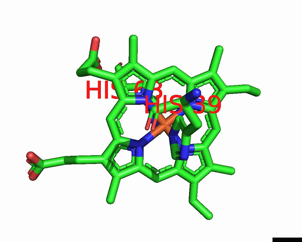
Mono view
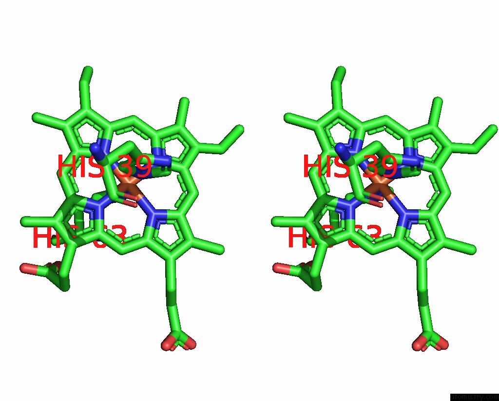
Stereo pair view
Mono view
Stereo pair view
A full contact list of Iron with other atoms in the Fe binding
site number 1 of Crystal Structure of Recombinant Trypsin-Solubilized Fragment of Cytochrome B5 within 5.0Å range:
|
Reference:
J.Wu,
J.H.Gan,
Z.X.Xia,
Y.H.Wang,
W.H.Wang,
L.L.Xue,
Y.Xie,
Z.X.Huang.
Crystal Structure of Recombinant Trypsin-Solubilized Fragment of Cytochrome B(5) and the Structural Comparison with VAL61HIS Mutant. Proteins V. 40 249 2000.
ISSN: ISSN 0887-3585
PubMed: 10842340
DOI: 10.1002/(SICI)1097-0134(20000801)40:2<249::AID-PROT70>3.0.CO;2-H
Page generated: Wed Jul 16 13:48:51 2025
ISSN: ISSN 0887-3585
PubMed: 10842340
DOI: 10.1002/(SICI)1097-0134(20000801)40:2<249::AID-PROT70>3.0.CO;2-H
Last articles
Fe in 2YXOFe in 2YRS
Fe in 2YXC
Fe in 2YNM
Fe in 2YVJ
Fe in 2YP1
Fe in 2YU2
Fe in 2YU1
Fe in 2YQB
Fe in 2YOO