Iron »
PDB 1eb7-1esz »
1eir »
Iron in PDB 1eir: 2,3-Dihydroxybiphenyl-1,2-Dioxygenase
Enzymatic activity of 2,3-Dihydroxybiphenyl-1,2-Dioxygenase
All present enzymatic activity of 2,3-Dihydroxybiphenyl-1,2-Dioxygenase:
1.13.11.39;
1.13.11.39;
Protein crystallography data
The structure of 2,3-Dihydroxybiphenyl-1,2-Dioxygenase, PDB code: 1eir
was solved by
T.Senda,
with X-Ray Crystallography technique. A brief refinement statistics is given in the table below:
| Resolution Low / High (Å) | 60.00 / 2.00 |
| Space group | I 4 2 2 |
| Cell size a, b, c (Å), α, β, γ (°) | 121.600, 121.600, 108.700, 90.00, 90.00, 90.00 |
| R / Rfree (%) | 18.1 / 23.1 |
Iron Binding Sites:
The binding sites of Iron atom in the 2,3-Dihydroxybiphenyl-1,2-Dioxygenase
(pdb code 1eir). This binding sites where shown within
5.0 Angstroms radius around Iron atom.
In total only one binding site of Iron was determined in the 2,3-Dihydroxybiphenyl-1,2-Dioxygenase, PDB code: 1eir:
In total only one binding site of Iron was determined in the 2,3-Dihydroxybiphenyl-1,2-Dioxygenase, PDB code: 1eir:
Iron binding site 1 out of 1 in 1eir
Go back to
Iron binding site 1 out
of 1 in the 2,3-Dihydroxybiphenyl-1,2-Dioxygenase
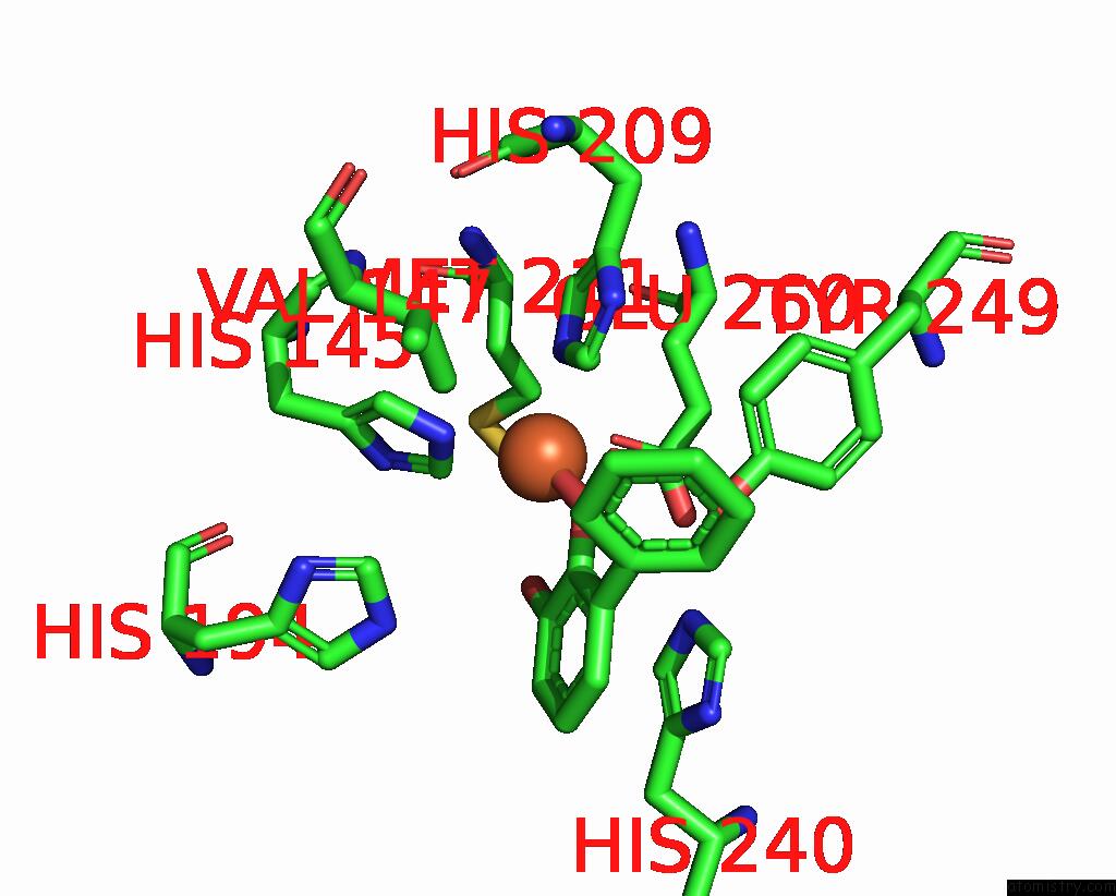
Mono view
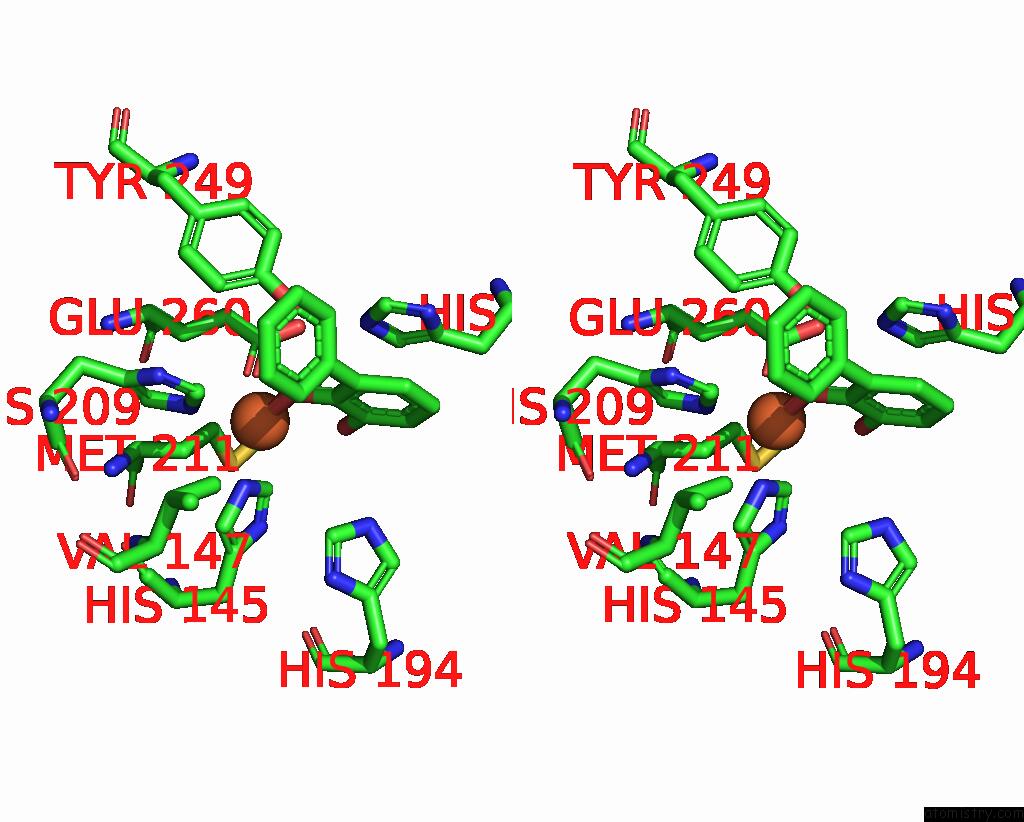
Stereo pair view
Mono view
Stereo pair view
A full contact list of Iron with other atoms in the Fe binding
site number 1 of 2,3-Dihydroxybiphenyl-1,2-Dioxygenase within 5.0Å range:
|
Reference:
Y.Uragami,
T.Senda,
K.Sugimoto,
N.Sato,
V.Nagarajan,
E.Masai,
M.Fukuda,
Y.Mitsu.
Crystal Structures of Substrate Free and Complex Forms of Reactivated Bphc, An Extradiol Type Ring-Cleavage Dioxygenase. J.Inorg.Biochem. V. 83 269 2001.
ISSN: ISSN 0162-0134
PubMed: 11293547
DOI: 10.1016/S0162-0134(00)00172-0
Page generated: Wed Jul 16 13:49:37 2025
ISSN: ISSN 0162-0134
PubMed: 11293547
DOI: 10.1016/S0162-0134(00)00172-0
Last articles
Fe in 2YXOFe in 2YRS
Fe in 2YXC
Fe in 2YNM
Fe in 2YVJ
Fe in 2YP1
Fe in 2YU2
Fe in 2YU1
Fe in 2YQB
Fe in 2YOO