Iron »
PDB 1eb7-1esz »
1eo2 »
Iron in PDB 1eo2: Crystal Structure of Acinetobacter Sp. ADP1 Protocatechuate 3,4- Dioxygenase
Enzymatic activity of Crystal Structure of Acinetobacter Sp. ADP1 Protocatechuate 3,4- Dioxygenase
All present enzymatic activity of Crystal Structure of Acinetobacter Sp. ADP1 Protocatechuate 3,4- Dioxygenase:
1.13.11.3;
1.13.11.3;
Protein crystallography data
The structure of Crystal Structure of Acinetobacter Sp. ADP1 Protocatechuate 3,4- Dioxygenase, PDB code: 1eo2
was solved by
M.W.Vetting,
D.A.D'argenio,
L.N.Ornston,
D.H.Ohlendorf,
with X-Ray Crystallography technique. A brief refinement statistics is given in the table below:
| Resolution Low / High (Å) | 20.00 / 2.25 |
| Space group | I 2 3 |
| Cell size a, b, c (Å), α, β, γ (°) | 145.500, 145.500, 145.500, 90.00, 90.00, 90.00 |
| R / Rfree (%) | 17.6 / 21.8 |
Iron Binding Sites:
The binding sites of Iron atom in the Crystal Structure of Acinetobacter Sp. ADP1 Protocatechuate 3,4- Dioxygenase
(pdb code 1eo2). This binding sites where shown within
5.0 Angstroms radius around Iron atom.
In total only one binding site of Iron was determined in the Crystal Structure of Acinetobacter Sp. ADP1 Protocatechuate 3,4- Dioxygenase, PDB code: 1eo2:
In total only one binding site of Iron was determined in the Crystal Structure of Acinetobacter Sp. ADP1 Protocatechuate 3,4- Dioxygenase, PDB code: 1eo2:
Iron binding site 1 out of 1 in 1eo2
Go back to
Iron binding site 1 out
of 1 in the Crystal Structure of Acinetobacter Sp. ADP1 Protocatechuate 3,4- Dioxygenase
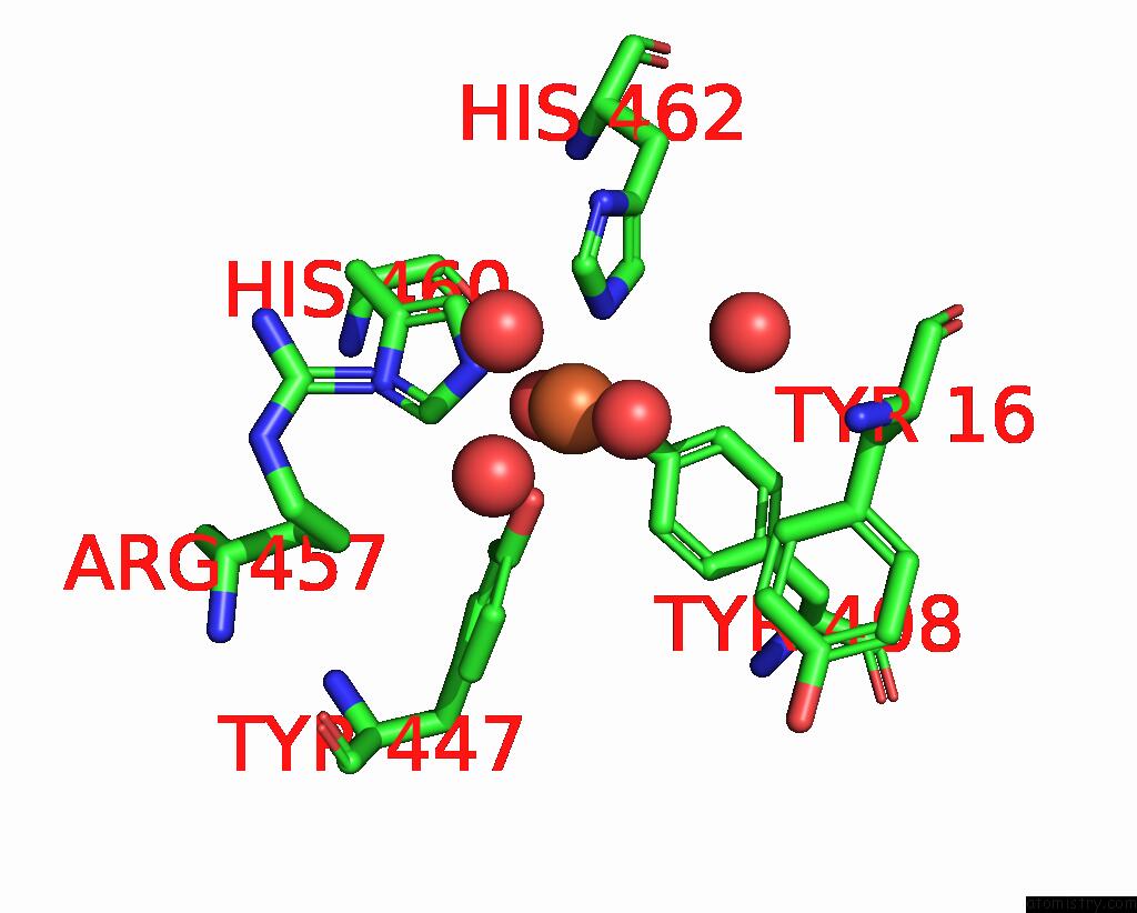
Mono view
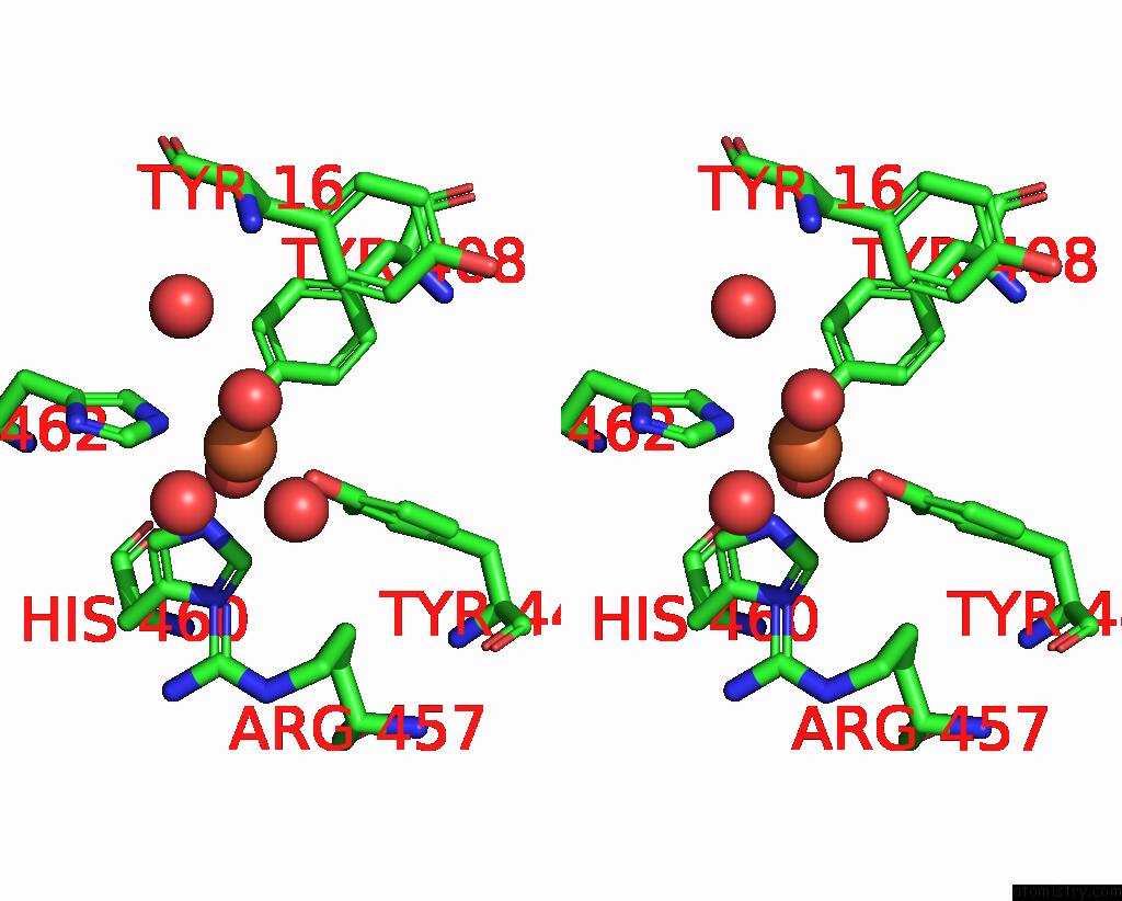
Stereo pair view
Mono view
Stereo pair view
A full contact list of Iron with other atoms in the Fe binding
site number 1 of Crystal Structure of Acinetobacter Sp. ADP1 Protocatechuate 3,4- Dioxygenase within 5.0Å range:
|
Reference:
M.W.Vetting,
D.A.D'argenio,
L.N.Ornston,
D.H.Ohlendorf.
Structure of Acinetobacter Strain ADP1 Protocatechuate 3, 4-Dioxygenase at 2.2 A Resolution: Implications For the Mechanism of An Intradiol Dioxygenase. Biochemistry V. 39 7943 2000.
ISSN: ISSN 0006-2960
PubMed: 10891075
DOI: 10.1021/BI000151E
Page generated: Wed Jul 16 13:49:53 2025
ISSN: ISSN 0006-2960
PubMed: 10891075
DOI: 10.1021/BI000151E
Last articles
Fe in 2YXOFe in 2YRS
Fe in 2YXC
Fe in 2YNM
Fe in 2YVJ
Fe in 2YP1
Fe in 2YU2
Fe in 2YU1
Fe in 2YQB
Fe in 2YOO