Iron »
PDB 1etp-1faw »
1eup »
Iron in PDB 1eup: X-Ray Crystal Structure of Cytochrome P450ERYF with Androstendione Bound
Protein crystallography data
The structure of X-Ray Crystal Structure of Cytochrome P450ERYF with Androstendione Bound, PDB code: 1eup
was solved by
J.R.Cupp-Vickery,
R.Anderson,
Z.Hatziris,
with X-Ray Crystallography technique. A brief refinement statistics is given in the table below:
| Resolution Low / High (Å) | 10.00 / 2.10 |
| Space group | P 21 21 21 |
| Cell size a, b, c (Å), α, β, γ (°) | 54.154, 79.114, 99.430, 90.00, 90.00, 90.00 |
| R / Rfree (%) | 18.4 / n/a |
Iron Binding Sites:
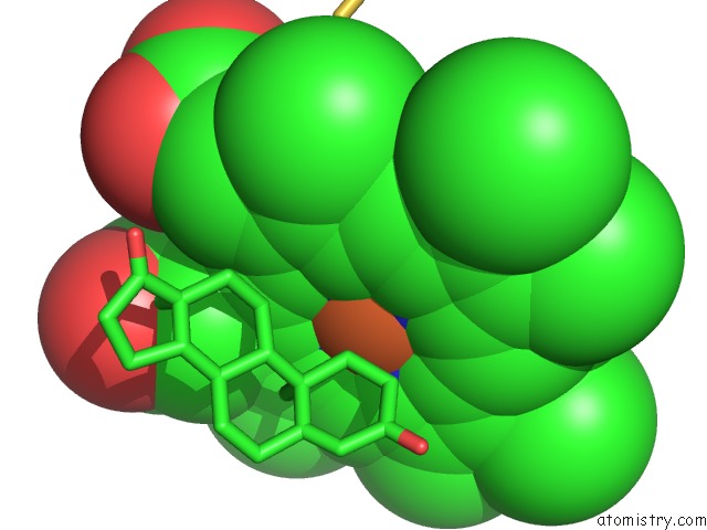
The binding sites of Iron atom in the X-Ray Crystal Structure of Cytochrome P450ERYF with Androstendione Bound
(pdb code 1eup). This binding sites where shown within
5.0 Angstroms radius around Iron atom.
In total only one binding site of Iron was determined in the X-Ray Crystal Structure of Cytochrome P450ERYF with Androstendione Bound, PDB code: 1eup:
In total only one binding site of Iron was determined in the X-Ray Crystal Structure of Cytochrome P450ERYF with Androstendione Bound, PDB code: 1eup:
Iron binding site 1 out of 1 in 1eup
Go back to
Iron binding site 1 out
of 1 in the X-Ray Crystal Structure of Cytochrome P450ERYF with Androstendione Bound
Mono view
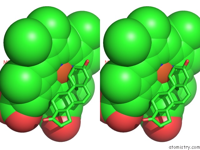
Stereo pair view
Mono view
Stereo pair view
A full contact list of Iron with other atoms in the Fe binding
site number 1 of X-Ray Crystal Structure of Cytochrome P450ERYF with Androstendione Bound within 5.0Å range:
|
Reference:
J.Cupp-Vickery,
R.Anderson,
Z.Hatziris.
Crystal Structures of Ligand Complexes of P450ERYF Exhibiting Homotropic Cooperativity. Proc.Natl.Acad.Sci.Usa V. 97 3050 2000.
ISSN: ISSN 0027-8424
PubMed: 10716705
DOI: 10.1073/PNAS.050406897
Page generated: Wed Jul 16 13:51:38 2025
ISSN: ISSN 0027-8424
PubMed: 10716705
DOI: 10.1073/PNAS.050406897
Last articles
Mg in 7FS1Mg in 7FS0
Mg in 7FRZ
Mg in 7FRY
Mg in 7FRX
Mg in 7FRW
Mg in 7FRV
Mg in 7FJP
Mg in 7FQJ
Mg in 7FGI