Iron »
PDB 1fca-1fnp »
1fhj »
Iron in PDB 1fhj: Crystal Structure of Aquomet Hemoglobin-I of the Maned Wolf (Chrysocyon Brachyurus) at 2.0 Resolution.
Protein crystallography data
The structure of Crystal Structure of Aquomet Hemoglobin-I of the Maned Wolf (Chrysocyon Brachyurus) at 2.0 Resolution., PDB code: 1fhj
was solved by
V.Fadel,
W.F.De Azevedo,
with X-Ray Crystallography technique. A brief refinement statistics is given in the table below:
| Resolution Low / High (Å) | 10.00 / 1.80 |
| Space group | P 21 21 21 |
| Cell size a, b, c (Å), α, β, γ (°) | 54.184, 87.725, 133.450, 90.00, 90.00, 90.00 |
| R / Rfree (%) | 19.4 / 24.6 |
Iron Binding Sites:
The binding sites of Iron atom in the Crystal Structure of Aquomet Hemoglobin-I of the Maned Wolf (Chrysocyon Brachyurus) at 2.0 Resolution.
(pdb code 1fhj). This binding sites where shown within
5.0 Angstroms radius around Iron atom.
In total 4 binding sites of Iron where determined in the Crystal Structure of Aquomet Hemoglobin-I of the Maned Wolf (Chrysocyon Brachyurus) at 2.0 Resolution., PDB code: 1fhj:
Jump to Iron binding site number: 1; 2; 3; 4;
In total 4 binding sites of Iron where determined in the Crystal Structure of Aquomet Hemoglobin-I of the Maned Wolf (Chrysocyon Brachyurus) at 2.0 Resolution., PDB code: 1fhj:
Jump to Iron binding site number: 1; 2; 3; 4;
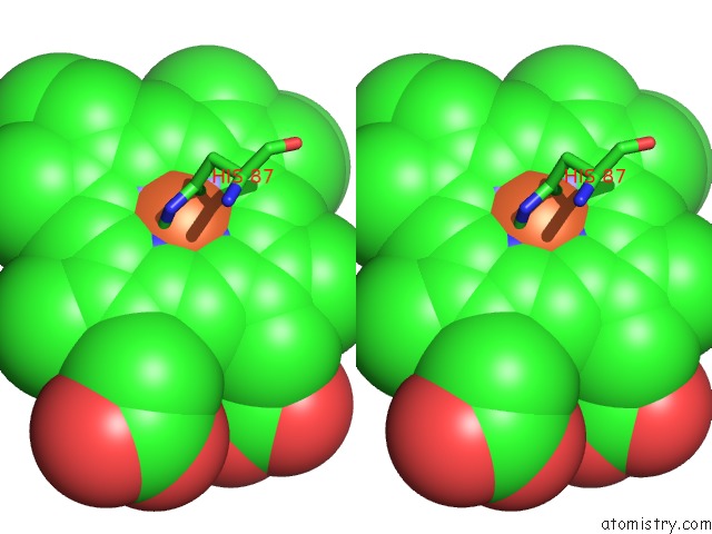
Iron binding site 1 out of 4 in 1fhj
Go back to
Iron binding site 1 out
of 4 in the Crystal Structure of Aquomet Hemoglobin-I of the Maned Wolf (Chrysocyon Brachyurus) at 2.0 Resolution.
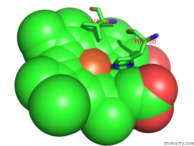
Mono view
Stereo pair view
Mono view
Stereo pair view
A full contact list of Iron with other atoms in the Fe binding
site number 1 of Crystal Structure of Aquomet Hemoglobin-I of the Maned Wolf (Chrysocyon Brachyurus) at 2.0 Resolution. within 5.0Å range:
|
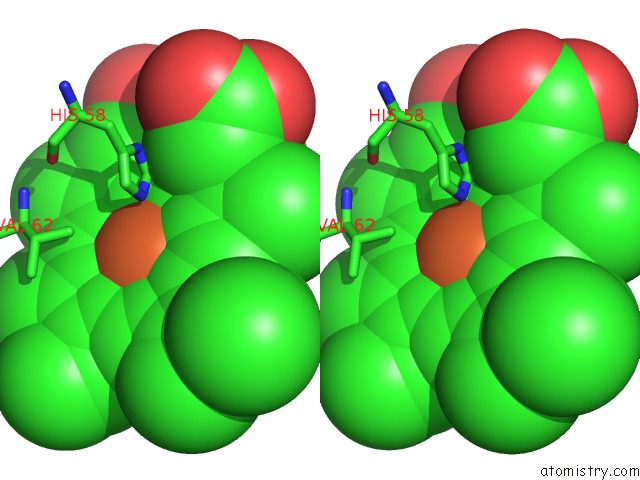
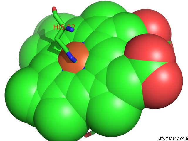
Iron binding site 2 out of 4 in 1fhj
Go back to
Iron binding site 2 out
of 4 in the Crystal Structure of Aquomet Hemoglobin-I of the Maned Wolf (Chrysocyon Brachyurus) at 2.0 Resolution.

Mono view
Stereo pair view
Mono view
Stereo pair view
A full contact list of Iron with other atoms in the Fe binding
site number 2 of Crystal Structure of Aquomet Hemoglobin-I of the Maned Wolf (Chrysocyon Brachyurus) at 2.0 Resolution. within 5.0Å range:
|
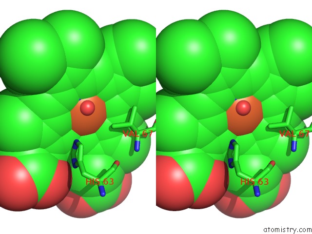
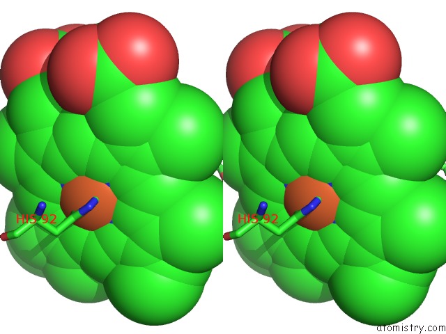
Iron binding site 3 out of 4 in 1fhj
Go back to
Iron binding site 3 out
of 4 in the Crystal Structure of Aquomet Hemoglobin-I of the Maned Wolf (Chrysocyon Brachyurus) at 2.0 Resolution.
Mono view
Stereo pair view
Mono view
Stereo pair view
A full contact list of Iron with other atoms in the Fe binding
site number 3 of Crystal Structure of Aquomet Hemoglobin-I of the Maned Wolf (Chrysocyon Brachyurus) at 2.0 Resolution. within 5.0Å range:
|
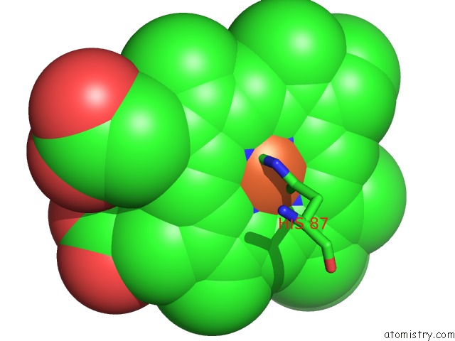
Iron binding site 4 out of 4 in 1fhj
Go back to
Iron binding site 4 out
of 4 in the Crystal Structure of Aquomet Hemoglobin-I of the Maned Wolf (Chrysocyon Brachyurus) at 2.0 Resolution.
Mono view
Stereo pair view
Mono view
Stereo pair view
A full contact list of Iron with other atoms in the Fe binding
site number 4 of Crystal Structure of Aquomet Hemoglobin-I of the Maned Wolf (Chrysocyon Brachyurus) at 2.0 Resolution. within 5.0Å range:
|
Reference:
V.Fadel,
F.Canduri,
J.R.Olivieri,
A.L.Smarra,
M.F.Colombo,
G.O.Bonilla-Rodriguez,
W.F.De Azevedo.
Crystal Structure of Hemoglobin From the Maned Wolf (Chrysocyon Brachyurus) Using Synchrotron Radiation. Protein Pept.Lett. V. 10 551 2003.
ISSN: ISSN 0929-8665
PubMed: 14683506
Page generated: Wed Jul 16 14:13:27 2025
ISSN: ISSN 0929-8665
PubMed: 14683506
Last articles
Pt in 5LXWPt in 5L3I
Pt in 5L3H
Pt in 5L75
Pt in 5K08
Pt in 5HMV
Pt in 5ILF
Pt in 5ILC
Pt in 5IDD
Pt in 5II3